
Table of Contents
When Your Skin Won’t Stop Building: What Abnormal Keratinocyte Proliferation Really Means
From stubborn scaly patches to sun-damage warning signs, a plain guide to skin cells that have stopped following the schedule
Picture a retired mason sitting in a dermatology clinic with what he calls “a dry patch” on his scalp. It’s been there about two years. He has tried coconut oil, a medicated shampoo a neighbor swore by, and, on one bad week, a steel comb to scrape it off. The patch keeps coming back, a little thicker each time. It turns out to be an actinic keratosis, and it’s the first of fourteen his dermatologist finds that afternoon.
Now picture someone completely different: a college student whose elbows have been scaly since exam season, tired of explaining that no, it isn’t catching. Two people, two patches, one shared problem. The skin cells involved have stopped following the schedule.
That problem has a clinical name, abnormal keratinocyte proliferation, and it sits underneath some of the most common skin conditions on earth: psoriasis, chronic eczema, sun-damage lesions, warts, and the skin cancers that rank among the most frequent cancers in people. This guide walks through what keratinocytes do, what goes wrong, how doctors sort harmless from dangerous, and what you can realistically do about it. Where a medical term matters, you’ll get the plain-English version first.

Meet the Keratinocyte: The Cell Doing Most of the Work
Around nine in every ten cells in your epidermis are keratinocytes. Melanocytes get the attention because of pigment, and Langerhans cells because of immunity, but the keratinocyte is the workhorse. It builds the barrier that keeps water in and germs out, and it does that by manufacturing keratin, the same tough protein family found in hair and nails.
Think of the epidermis as a brick wall that rebuilds itself from the foundation up while the top row is constantly swept away. New cells form at the bottom. They climb, change shape, fill with keratin, and eventually become flat, dead, protective plates that flake off without anyone noticing.
Skin cell turnover: the normal timetable
In healthy skin, a keratinocyte takes roughly four weeks to travel from the bottom layer to the surface and shed. It slows a bit with age, which is partly why older skin heals more slowly. That month-long journey matters, because every stage of it is a chance for the cell to mature properly. This maturation process is called epidermal differentiation, and when it goes wrong, the skin goes wrong with it.
The four floors that matter
| Layer | What normally happens | What goes wrong with overgrowth |
| Stratum basale (bottom) | Stem-like keratinocytes divide to supply new cells | Too many divisions; the living epidermis thickens (acanthosis) |
| Stratum spinosum | Cells link together and produce keratin | Swelling between cells in eczema; crowding in psoriasis |
| Stratum granulosum | Barrier proteins such as filaggrin are assembled | Often thin or missing in psoriasis; weak in atopic skin |
| Stratum corneum (surface) | Flat, dead cells form the waterproof shield | Excess buildup (hyperkeratosis) or cells that keep their nuclei (parakeratosis) |
Palms and soles add a clear fifth layer, the stratum lucidum, because those areas need extra armor. Everywhere else, the four floors above do the job.
Accelerators and brakes
Every cell-division system needs both. In keratinocytes, the accelerators include growth factors such as TGF-alpha and other members of the EGF family, plus inflammatory messengers like IL-17, IL-22 and IL-23. The brakes include the p53 protein, which pauses damaged cells so they can be repaired (or sends them off to self-destruct), the retinoblastoma protein, Notch signaling, and the calcium gradient that tells a cell it’s time to mature instead of multiply.
That gives you a clean way to think about keratinocyte hyperproliferation: an accelerator stuck down, a brake worn out, or both. The reason it shows up in such different diseases is that the cause of the stuck pedal differs:
- Immune signals keep pressing the accelerator (psoriasis)
- Sun damage wears out the brakes by mutating p53 (actinic keratosis, squamous cell carcinoma)
- Viral hijacking overrides the brakes from the outside (warts caused by HPV)
- Barrier failure and constant scratching keep the skin stuck in repair mode (chronic eczema)
What “Abnormal” Actually Means
Here’s something that surprises people: thick skin isn’t automatically a problem. The callus on a cobbler’s thumb is keratinocytes proliferating, and it’s doing exactly what it should. Skin responds to friction and pressure by building more of itself. Doctors call this reactive hyperplasia, and it fades once the trigger goes away.
Wound healing is the other everyday example. Within hours of a cut, keratinocytes at the edge switch into a migratory mode, crawl across the wound bed, and multiply to seal it. Then they switch back. That on-and-off discipline is the whole point. Abnormal proliferation is what you get when the “off” switch never fully flips.
Abnormal proliferation is different. The growth is out of proportion to any need, it persists without a trigger, or the cells themselves have changed. Pathologists describe it along a spectrum, and where a patch sits on that spectrum decides almost everything about what happens next. Simple epidermal hyperplasia, where there are more cells but they’re orderly, sits at the mild end.
A traffic analogy that works
Picture a highway. Hyperplasia is rush hour: heavy traffic, but everyone stays in lane and obeys the signals. Dysplasia is the stretch where some drivers start swerving and ignoring the markings. The cells look atypical and the layering is disordered. Carcinoma in situ is when the swerving drivers are all over the road but still haven’t left it. Invasive carcinoma is when they’ve gone through the guardrail and into the neighborhood below, which in skin means crossing the basement membrane into the dermis.
The vocabulary on your biopsy report
| Term | Plain-English meaning | Commonly seen in |
| Acanthosis | A thickened living layer of epidermis | Psoriasis, eczema, warts |
| Hyperkeratosis | Too much buildup of the dead surface layer | Calluses, actinic keratosis |
| Parakeratosis | Surface cells that kept their nuclei because they were rushed upward | Psoriasis, actinic keratosis |
| Dysplasia | Atypical, disorganized cells | Actinic keratosis |
| Carcinoma in situ | Malignant-looking cells still confined to the epidermis | Bowen disease |
| Invasive carcinoma | Cancer cells that have broken through into the dermis | Squamous cell carcinoma |
The real question is never just “are there too many cells?” It’s “are they still orderly, and are they still listening to the brakes?”
The Conditions Behind the Overgrowth
Abnormal keratinocyte proliferation is a mechanism, not a diagnosis. These are the conditions where you’re most likely to meet it.
Psoriasis: when keratinocytes join the immune loop
Psoriasis affects roughly two to three percent of people worldwide, and the keratinocyte is both victim and accomplice. It starts with immune cells. Dendritic cells release IL-23, which prompts T helper 17 cells to produce IL-17 and IL-22. Those signals tell keratinocytes to multiply at enormous speed, and the stressed cells respond by sending out chemical flares that recruit even more immune cells. That’s the loop. Psoriasis keratinocytes don’t just react to inflammation, they feed it.
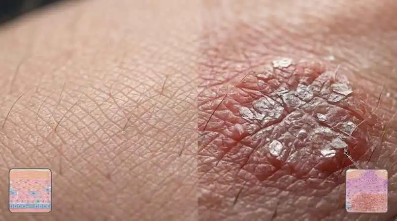
The turnover numbers show how dramatic this is. Where a normal cell takes about a month to reach the surface, a psoriatic one can make the trip in just a few days. The cells arrive unfinished, which is why you see thick silvery scale on red, sharply defined plaques, most often on the elbows, knees, scalp and lower back. Psoriasis isn’t contagious, and it has nothing to do with hygiene. It’s also more than skin: a meaningful share of patients develop psoriatic arthritis, and the chronic inflammation is linked with cardiovascular and metabolic risk.
Chronic eczema and the leathery skin problem
Atopic dermatitis starts from a different place. Often the barrier itself is the weak point, sometimes through changes in the filaggrin gene, so poor skin barrier function lets irritants and allergens in and triggers a different immune pathway involving IL-4 and IL-13. Itch leads to scratching, scratching injures keratinocytes, and injured keratinocytes release more inflammatory signals. Years of this cycle can thicken the skin into leathery, exaggerated-crease patches called lichenification. That’s abnormal proliferation by attrition, powered by the hand rather than the gene.
Actinic keratosis: the sun-damage warning sign
Actinic keratoses are rough, dry, sandpaper-like spots on skin that has had decades of sun: scalp, forehead, ears, nose, backs of the hands, forearms, and lower lip. Sometimes you feel them before you see them. Ultraviolet radiation causes mutations in the p53 gene, and keratinocytes carrying those mutations keep dividing when they should have been stopped.
Most individual lesions never become cancer. Some regress, and some sit unchanged for years. The catch is that nobody can look at one and predict which way it will go. And because the surrounding skin carries the same damage (dermatologists call this field cancerization), treatment usually targets the whole field, not just the visible spot.
Squamous cell carcinoma and basal cell carcinoma
Squamous cell carcinoma arises from keratinocytes higher in the epidermis, and basal cell carcinoma from cells in the basal layer. Together they’re among the most common cancers in people. Basal cell carcinomas grow slowly and rarely spread, but they can destroy local tissue. Squamous cell carcinomas are more likely to invade deeper and, less often, to reach the lymph nodes, particularly when they start on the lips or ears or in people with weakened immunity. Both are highly treatable when caught early.
Bowen disease deserves a mention here. It’s squamous cell carcinoma still confined to the epidermis, and it often looks like a persistent scaly red patch that gets mistaken for eczema or psoriasis.
The benign and the borrowed
Not every proliferation is dangerous. Seborrheic keratosis, those waxy, stuck-on, brown-to-black growths that multiply with age, is a benign overgrowth of keratinocytes. It’s harmless, though it can occasionally mimic melanoma and is worth showing a doctor if one changes. Warts are another case: human papillomavirus makes proteins (E6 and E7) that switch off the cell’s own brakes, so the infected keratinocyte keeps dividing and the virus gets a bigger home. Some inherited disorders, including certain ichthyoses and pityriasis rubra pilaris, also feature disordered growth and keratinization.
| Condition | Main driver | Typical look | Cancer concern |
| Psoriasis | IL-23/IL-17 immune loop | Thick silvery scale on red plaques | Not cancerous |
| Chronic atopic dermatitis | Barrier defect, inflammation, scratching | Dry, itchy, thickened leathery patches | Not cancerous |
| Actinic keratosis | UV-damaged p53 | Rough sandpaper patches on sun-exposed skin | Possible precursor to SCC |
| Squamous cell carcinoma | UV, immunosuppression, HPV | Firm, scaly or crusted nodule that may bleed | Malignant; can spread |
| Basal cell carcinoma | UV, hedgehog pathway mutations | Pearly bump with tiny vessels, or a non-healing sore | Malignant; rarely spreads |
| Seborrheic keratosis | Age-related, benign | Waxy, stuck-on brown growth | Benign |
| Viral wart | HPV disabling cell brakes | Rough raised bump, sometimes with dark dots | Usually benign |
Spotting It Early: Signs, Risk Factors and How Doctors Confirm
Signs worth noticing
Your skin tells you when growth is going sideways, if you know what to look for. Pay attention to:
- A scaly patch that returns after every round of moisturizer, always in the same spot
- A rough area you feel more than you see
- A sore that doesn’t heal within about four weeks, or one that heals and reopens
- A lump that’s firm, growing, tender, or bleeds with the slightest contact
- A red, scaly patch that doesn’t respond to steroid or antifungal cream the way you’d expect
- Thick silvery scale together with pitted or crumbling nails
- Any lesion that looks different from its neighbors
The ABCDE rule you may have heard of is built for melanoma. For keratinocyte-related lesions a rougher rule of thumb works better: if it’s scaly, bleeding or not healing, and it’s been there longer than a month, get it checked.
Who is at higher risk
- People with fair skin, light eyes, or a tendency to burn rather than tan
- Outdoor workers: farmers, fishers, construction crews, road workers, delivery riders, anyone with decades of daily sun
- Adults over 50, though cumulative sun exposure matters more than the birthday itself
- Organ transplant recipients and others on long-term immunosuppressive medicine, who face dramatically higher squamous cell risk
- Anyone with a history of tanning-bed use, severe sunburns, or a previous skin cancer
- People with a family history of psoriasis (for the inflammatory conditions) or skin cancer
Deeper skin tones lower the risk of sun-driven lesions but don’t erase it. When cancers do appear in darker skin, they’re more often found in places like old scars, chronic wounds and the legs, and they’re frequently diagnosed late because nobody was looking there.
How doctors confirm it
- History and exam. How long it’s been there, how it has changed, what you’ve tried, your sun history and medications.
- A handheld magnifier with polarized light shows vessel patterns and surface structures the naked eye misses.
- A small sample, taken by shave, punch or excision, goes to a pathologist. For anything suspicious, this is the definitive step.
- The pathologist checks layer thickness, nuclear atypia, whether cells are orderly, and whether anything has crossed the basement membrane.
- Staging when needed. High-risk squamous cell cancers may call for a lymph node exam or imaging.
Questions worth bringing to your appointment
- What do you think this is, and what else could it be?
- Do you recommend a biopsy, and what will it tell us?
- Is this a single lesion, or part of a larger sun-damaged field?
- What are my options, and what will each one look like day to day?
- How long until I should see a change, and what should make me call sooner?
- When should I come back for a follow-up skin check?
Myths that keep people from getting help
| The myth | The reality |
| “It’s just dry skin. Moisturizer will fix it.” | Dry skin responds to moisturizer within days. A patch that keeps returning to the same spot needs a diagnosis. |
| “Skin cancer only happens to fair-skinned sunbathers.” | Anyone can develop it. Outdoor workers, people with weakened immunity, and people with darker skin (often at sites that rarely see sun) are all affected. |
| “If it doesn’t hurt, it isn’t serious.” | Many precancers and early skin cancers are painless. |
| “Psoriasis comes from poor hygiene or a bad diet.” | It’s an immune-driven, largely inherited condition. Hygiene has nothing to do with it. |
| “A scab that keeps forming just needs more air.” | A scab that forms, falls off and forms again in the same place is a classic sign of a non-healing lesion. |
| “Once it’s removed, it’s gone for good.” | The lesion is, but sun-damaged skin can grow new ones, which is why follow-up checks matter. |
Psoriasis is usually recognized by its look and pattern, and biopsy is kept for doubtful cases. One practical tip: if you can help it, don’t spend weeks putting steroid cream on a mystery patch before the appointment. It can blur the picture and make the biopsy harder to read.
Treatment Roadmap: From Creams to Biologics
Treatment depends on the diagnosis, and the diagnosis depends on looking properly, so the order matters: diagnose first, then treat. Here’s the toolkit.
Topical treatments
- Topical corticosteroids calm inflammation in psoriasis and eczema. Potency, body site and duration need care, because long use thins the skin.
- Vitamin D analogs such as calcipotriol slow keratinocyte division and help the cells mature normally.
- Calcineurin inhibitors (tacrolimus, pimecrolimus) are steroid-free options for the face and skin folds.
- Topical retinoids such as tazarotene nudge differentiation back toward normal.
- Keratolytics (salicylic acid, urea, lactic acid) soften and lift scale. They don’t stop overproduction.
- 5-fluorouracil, imiquimod, diclofenac and tirbanibulin are field therapies for actinic keratosis. They target abnormal cells, and the redness and crusting they cause over days to weeks is usually the sign they’re working.
Light-based treatment
Narrowband UVB phototherapy slows overactive keratinocytes and calms the T cells driving psoriasis, typically with two to three sessions a week over a few months. Photodynamic therapy takes a different route: a light-sensitizing medicine is applied to the skin, abnormal cells absorb more of it, and a specific light then destroys them selectively. It’s widely used for fields of actinic keratosis.
Systemic medicines and biologics
When disease is widespread, stubborn, or reaching the joints, doctors move beyond the skin. Older systemic options include methotrexate, cyclosporine and acitretin, a retinoid that normalizes keratinocyte growth. Newer ones include apremilast, deucravacitinib, and biologics that block TNF, IL-17, IL-23 or IL-12/23.
Biologics go after the signals that tell keratinocytes to overgrow rather than the keratinocytes themselves, and they’ve changed psoriasis care: many people reach clear or almost clear skin. They require screening (for tuberculosis and hepatitis, for instance) and monitoring, so these decisions belong with a dermatologist.
Procedures for individual lesions
- Cryotherapy freezes isolated actinic keratoses and warts with liquid nitrogen.
- Curettage and electrodesiccation scrape and cauterize certain superficial lesions.
- Surgical excision is standard for many squamous and basal cell carcinomas.
- Mohs micrographic surgery removes cancer layer by layer, checking each under a microscope during the operation, which preserves healthy tissue. It’s favored for the face, ears, and recurrent or high-risk tumors.
- Radiation therapy is an option when surgery isn’t practical.
A quick note on timing, because impatience wrecks more treatment plans than anything else. Topical psoriasis treatments usually need two to four weeks before the verdict. Biologics often take two to four months to reach their full effect. Field therapy for actinic keratosis involves a few weeks of application, then a further stretch of healing as the skin settles. Ask your doctor what “working” should look like at each milestone, and don’t abandon a plan in week one because the skin looks angrier before it looks better.
| Approach | Best for | Watch-outs |
| Topical steroids | Psoriasis and eczema flares | Skin thinning with long use |
| Vitamin D analogs | Mild to moderate psoriasis | Stay within recommended amounts |
| Field therapy (5-FU, imiquimod) | Many actinic keratoses across one area | Weeks of redness and crusting |
| Phototherapy | Widespread psoriasis, some eczema | Frequent visits; cumulative UV exposure |
| Biologics | Moderate to severe psoriasis | Infection screening, cost, long-term monitoring |
| Mohs surgery | Facial or high-risk skin cancers | Needs a specialist; repair of the wound afterward |
Putting It Into Practice: Three Everyday Scenarios
Theory is tidy. Skin isn’t. Here’s how the thinking above plays out in three ordinary situations.
The farmer with a crusty spot on his ear
A man in his late fifties who has worked fields since his teens notices a crusty, slightly tender spot on the rim of his ear. It bleeds when he towels off, scabs over, and comes back. This is the classic profile of a keratinocyte lesion: decades of cumulative UV, an exposed site that sunscreen rarely reaches, and a sore that won’t stay healed. The sensible move is a prompt skin exam and probably a biopsy, since ears and lips are higher-risk sites for squamous cell carcinoma. Waiting for it to “dry out by itself” is exactly how a manageable problem turns into a surgical one.
The shopkeeper with thick, itchy shins
A woman who stands behind a counter all day develops dry, itchy patches on her shins that have gradually turned thick and leathery from night-time scratching. Nothing here points to cancer, and the pattern fits chronic eczema with lichenification. The first steps are unglamorous: lukewarm showers, a thick fragrance-free moisturizer within minutes of bathing, short nails, and a doctor-prescribed anti-inflammatory cream for the flare itself. Breaking the itch-and-scratch loop thins the skin back down far more reliably than any scrub.
The new father with scaly elbows
A young father develops silvery, sharply defined plaques on both elbows a few weeks after a throat infection and a run of sleepless nights. Infection and stress are both classic psoriasis triggers, and the symmetry and silvery scale fit. He doesn’t need to panic. He needs a diagnosis, a starting plan (often a steroid and vitamin D combination), and a conversation about morning joint stiffness, which can be an early sign of psoriatic arthritis.
Notice what the three have in common. Each person spotted a pattern, refused to dismiss it, and matched the next step to the likely mechanism. That’s really all this article is asking of you.
Daily Habits That Keep Keratinocytes in Line
Medicine handles the diagnosis. Habits handle the other three hundred and sixty days of the year.
Make sun protection boring and automatic
- Use a broad-spectrum sunscreen of SPF 30 or higher on exposed skin, apply it about 15 minutes before going out, and reapply every two hours outdoors
- Wear a wide-brim hat, a neck cover and long sleeves if your job keeps you outside. A hat costs far less than any treatment on this page.
- Seek shade during the strongest sun, roughly 10 a.m. to 4 p.m.
- Skip tanning beds entirely
Protect the barrier
A healthy barrier means fewer inflammatory signals. Moisturize right after bathing, choose fragrance-free creams, use lukewarm water, and avoid scrubbing. Don’t pick at scale. In psoriasis, injury can trigger new plaques exactly where the skin was hurt, a pattern called the Koebner phenomenon. (And yes, the steel comb was a bad idea.)
Know your triggers
Common culprits include stress, infections (strep throat can set off guttate psoriasis), skin injury, heavy alcohol use, smoking, weight gain, and certain medicines such as lithium and beta-blockers. A simple diary of flares and what happened beforehand will show patterns faster than guessing.
A five-minute monthly skin check
- Pick the same day each month, stand in good light, and have a full-length mirror and a hand mirror ready.
- Start at the top: scalp (part the hair with a comb, or ask a family member), face, ears and neck.
- Check the backs and palms of your hands, forearms, and between the fingers, then chest, belly and sides.
- Check legs, tops of feet, soles and between the toes.
- Photograph anything new or changing with a coin or ruler beside it for scale, and compare next month.
- Book an appointment for anything that bleeds, grows, hurts, or hasn’t healed in about four weeks.
A word on home remedies
Aloe, coconut oil, turmeric pastes and oatmeal baths can soothe dryness and itch, and plenty of people like them. None of them treats the underlying overgrowth, and none should stand in for a diagnosis on a lesion that might be precancerous. Delay is the real risk: a squamous cell cancer treated at six months is a very different problem from one treated at two years. Also, leave the scraping, cutting and burning to professionals. Doing it at home spreads infection, scars the skin, and can leave abnormal cells behind.
The Bottom Line
Abnormal keratinocyte proliferation sounds like a laboratory phrase, but it describes something you can see and feel: skin that builds faster than it should, or without listening to the usual stop signals. Sometimes the cause is an overactive immune system, sometimes decades of sun, sometimes a virus borrowing your cells. The encouraging part is that skin is one of the most visible and accessible organs in medicine. You can look at it. You can photograph it. You can show it to a specialist.
The mason from the start of this article isn’t a cautionary tale about carelessness. He’s a reminder that a patch you’ve been moisturizing for a year deserves a real answer.
Your next step: check your skin this week. Spend five minutes with a mirror, photograph anything unusual, and book a dermatology visit for any patch that’s scaly, sore or stubborn. If this guide helped, send it to someone who spends their days in the sun, and keep reading CloudTechBull for more clear, practical health explainers.
FAQs
What is abnormal keratinocyte proliferation in simple terms?
It means the main cells of your outer skin are multiplying faster than the body needs, or without responding to the normal stop signals. The result can be thickened, scaly or rough skin, and in some cases a growth that’s precancerous or cancerous. It’s a mechanism behind many conditions, not one single disease.
Is it the same thing as skin cancer?
No. Most abnormal proliferation isn’t cancer. Psoriasis, chronic eczema, warts and seborrheic keratoses all involve extra keratinocyte growth without being malignant. Skin cancers like squamous cell carcinoma sit at one end of the spectrum, which is why an unexplained, persistent patch deserves a proper look.
What causes keratinocytes to multiply too fast?
The main triggers are immune signals (psoriasis), ultraviolet damage to growth-control genes (actinic keratosis and squamous cell carcinoma), viral infection (HPV warts), chronic irritation or scratching (lichenified eczema), and inherited keratinization disorders. Often more than one factor stacks up, such as genetics plus an infection plus stress.
How fast does healthy skin renew itself?
A keratinocyte typically takes around four weeks to move from the basal layer to the surface and shed, though this slows with age. In psoriasis the journey can shrink to a few days, which is why immature cells pile up as thick scale.
Is psoriasis a skin problem or an immune problem?
Both, and they feed each other. Immune cells release IL-23, IL-17 and IL-22, which push keratinocytes to overgrow. The keratinocytes then release signals that recruit more immune cells. That’s why treatments aimed at the immune side, like biologics, can clear skin so effectively.
Is it contagious?
Psoriasis, eczema, actinic keratoses and skin cancers are not contagious. Warts are the exception. They’re caused by HPV and can spread through direct contact or damp shared surfaces such as shower floors, although not everyone exposed develops them.
Can stress make it worse?
Stress is a well-recognized flare trigger for psoriasis and eczema, through immune and hormonal pathways and through behavior like scratching or poor sleep. It doesn’t create the underlying tendency, but managing it can reduce how often flares show up.
Is an actinic keratosis a cancer?
It’s usually classed as a precancer, although some pathologists regard it as very early squamous cell carcinoma in situ. Either way, it consists of abnormal keratinocytes with sun-induced DNA damage. Doctors treat them because they can’t predict which ones will progress.
How often does an actinic keratosis turn into squamous cell carcinoma?
Published estimates vary widely, but the chance for any single lesion in a given year is generally low. The trouble is that a person with many lesions carries a higher overall risk, and nobody can reliably tell which spot will progress. That’s the reason treatment often covers the whole sun-damaged field.
What’s the difference between hyperplasia and dysplasia?
Hyperplasia means more cells in an orderly arrangement, like a callus. Dysplasia means the cells themselves look abnormal and the layering is disorganized. Hyperplasia is often reversible and benign, while dysplasia carries a greater chance of moving toward cancer.
Are seborrheic keratoses dangerous?
No, they’re benign. They’re common after about age 40, look waxy and stuck-on, and don’t turn into cancer. Have one examined if it bleeds, itches persistently, grows suddenly, or looks different from your others, since melanoma can occasionally imitate one.
Are warts a type of abnormal keratinocyte proliferation?
Yes. HPV proteins disable the cell’s growth brakes, so infected keratinocytes keep dividing and build the rough lump you see. Most common warts are harmless and clear on their own or with treatment, though persistent or unusual ones should be examined.
What does a skin biopsy actually show?
The pathologist examines the thickness of each layer, whether cells are orderly or atypical, how mature they are by the time they reach the surface, and whether any abnormal cells have crossed into the dermis. That tells your doctor whether you’re dealing with hyperplasia, dysplasia, in situ cancer or invasive cancer.
Can abnormal keratinocyte growth be cured?
It depends on the cause. Warts and individual actinic keratoses can be cleared, and early skin cancers are usually cured by removal. Psoriasis and atopic dermatitis are chronic conditions that can be controlled, sometimes to clear skin, but they tend to return if treatment stops.
Do moisturizers help?
They help with symptoms and the barrier: less scale, less itching, fewer cracks. They won’t stop the overgrowth on their own, and they won’t treat a precancerous lesion. Think of them as the foundation of care, not the treatment.
Does salicylic acid fix it?
It loosens and removes scale, which helps other treatments reach the skin, but it doesn’t slow cell production. Overuse irritates, and it shouldn’t be used on a suspicious lesion as a substitute for diagnosis.
Is sunscreen enough to prevent it?
Sunscreen matters, but it isn’t enough alone. Pair it with shade, hats, long sleeves and avoiding peak sun hours. Most people apply too little and don’t reapply, so real-world protection runs lower than the label suggests.
Do tanning beds raise the risk?
Yes. Tanning devices are classified as carcinogenic to humans, and using them is linked with a higher risk of squamous cell carcinoma, basal cell carcinoma and melanoma, especially when use starts young. There’s no such thing as a safe base tan.
Who is most at risk of sun-related keratinocyte cancers?
People with fair skin who burn easily, those with decades of outdoor exposure, adults over 50, anyone with many sunburns or tanning-bed use, people with a previous skin cancer, and anyone with a weakened immune system. Risk is lower in deeper skin tones, but it isn’t zero.
Why are transplant patients at higher risk?
The medicines that prevent organ rejection also suppress the immune surveillance that normally clears early abnormal cells. Squamous cell carcinoma risk can be dozens of times higher than in the general population, and the cancers can behave more aggressively. Regular full-skin exams are standard for these patients.
Does diet affect it?
No diet cures abnormal keratinocyte growth. In psoriasis, weight loss in people who are overweight can improve disease and treatment response, and heavy alcohol use tends to make it worse. Eating well supports general skin health, but supplements won’t replace medical treatment.
Does smoking matter?
Yes. Smoking is linked with higher psoriasis risk and more severe disease, and with squamous cell cancers of the lip and mouth. Quitting is one of the more useful steps a person with chronic skin disease can take.
Does scratching or picking make it worse?
It does. Injury can trigger new psoriasis plaques at the site (the Koebner phenomenon), and chronic scratching thickens eczema skin into leathery patches. Short nails, cool compresses and treating itch early all help break the cycle.
Are natural remedies like aloe or turmeric safe?
Many are fine for soothing mild dryness or itch, although some cause contact allergy. The bigger risk isn’t the remedy, it’s the delay. A lesion that might be precancerous needs a diagnosis first, and natural products don’t treat the underlying overgrowth.
What do biologics do for keratinocytes?
They don’t act on keratinocytes directly. They block messengers such as TNF, IL-17 or IL-23 that tell keratinocytes to multiply. With the signal gone, turnover normalizes and plaques fade. They require screening for infections like tuberculosis and ongoing monitoring.
Can children develop it?
For some conditions, yes. Psoriasis, atopic dermatitis and warts are common in childhood. Actinic keratoses and skin cancers are very rare in children, except in inherited conditions such as xeroderma pigmentosum, where cells can’t repair UV damage.
Is it hereditary?
Genes play a large role in psoriasis, atopic dermatitis and the inherited keratinization disorders, so these often run in families. Skin cancer risk reflects inherited traits like skin type combined with sun exposure over a lifetime.
How often should I have a skin check?
A monthly self-check, plus a clinician’s look whenever something seems off, suits people at average risk. With risk factors such as past skin cancer, many actinic keratoses or immunosuppression, dermatologists often recommend professional exams once or twice a year or more. Your doctor will set the schedule.
What does a sore that won’t heal usually mean?
Often it’s a minor injury or infection that needs better care. But a sore that fails to heal in about four weeks is a classic presentation of basal or squamous cell carcinoma, so it deserves a biopsy rather than another round of ointment.
When should I see a doctor urgently?
Seek prompt care for a fast-growing or bleeding lump, a painful ulcer, a new lump in the neck, armpit or groin near a skin lesion, or widespread red, peeling skin with fever or chills, which can signal erythrodermic psoriasis. Don’t wait for a routine appointment with any of those.
Medical Disclaimer : This article is for general education and isn’t a substitute for professional medical advice, diagnosis or treatment. See a qualified doctor or dermatologist about any skin change that concerns you.
