
Table of Contents
The Silent Disfigurement: Decoding the Medical Mystery of Hypopigmented Nodules and Nerve Thickening
In the sprawling, fast-paced world of medical social media, certain cases stop seasoned physicians and curious observers alike in their tracks. A recent case study of a 45-year-old male presenting with multiple, hypopigmented, entirely painless skin nodules, visibly thickened nerves, and profound, debilitating deformities of the hands and face.
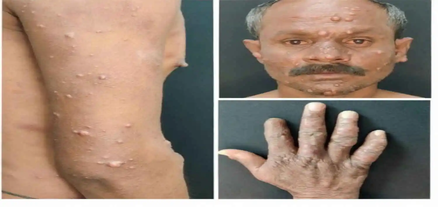
The image of the person was a stark documentation of a body slowly betraying itself. The arm displayed scattered, pale lesions that seemed to blend into the skin, devoid of sensation. The face showed deepened furrows and nodular thickenings that altered the patient’s natural features. But it was the image of the hands that revealed the true devastation—a claw-like retraction of the fingers, a classic physical testament to irreversible nerve damage.
With nearly a thousand likes and hundreds of frantic comments guessing everything from autoimmune disorders to rare genetic syndromes, its simple but daunting question: “Diagnosis? Treatment?”
This is not just a medical puzzle for doctors; it is a profound human story about a disease that has haunted humanity for millennia. To understand this case, we must delve deep into the pathogenesis, the devastating clinical progression, and the modern medical strategies used to combat one of the world’s oldest and most misunderstood diseases: Leprosy, specifically the Lucio Phenomenon and its associated Type 2 (Erythema Nodosum Leprosum) reactions, complicated by severe neuropathy.

The Clinical Picture: Deconstructing the Symptoms
To the untrained eye, the symptoms presented in the image might seem disconnected. However, to a dermatologist or a neurologist, they weave a highly specific tapestry. Let us break down the triad of symptoms presented by this 45-year-old patient.
- Multiple Hypopigmented, Painless Skin Nodules: The skin is the most visible window into this disease. The lesions described are “hypopigmented”—meaning they are lighter than the surrounding normal skin. This occurs because the underlying pathology directly targets the melanocytes (the cells responsible for skin pigment) and the skin’s immune architecture. Crucially, these lesions are “painless.” In most bacterial infections, pain is a primary symptom due to acute inflammation. The absence of pain in these nodules is a massive clinical red flag. It indicates that the sensory nerves supplying that specific patch of skin have already been destroyed or severely damaged. The nodular nature of the lesions suggests a high bacterial load (multibacillary disease) and a robust, albeit misplaced, immune response.
- Nerve Thickening: This is the absolute hallmark of the disease. Unlike most bacteria that wander through the bloodstream, the pathogen responsible for this condition—Mycobacterium leprae—has a highly specific thermal preference. It thrives at temperatures slightly below the core body temperature (around 27°C to 30°C). Because of this, it does not invade the internal organs; instead, it aggressively attacks the coolest parts of the body: the skin, superficial nerves, testes, and eyes.
The bacteria invade Schwann cells, the protective wrapping around peripheral nerves. As the body’s immune system attempts to fight the infection, the nerves become inflamed, swollen, and thickened. A doctor can often physically feel these enlarged nerves rolling under the skin, particularly around the elbows (ulnar nerve), knees (peroneal nerve), and neck (great auricular nerve).
- Hand and Facial Deformities: The deformities seen in the photographs are not caused by the bacteria directly destroying the bone or muscle. They are an indirect, tragic consequence of the nerve damage. When the sensory nerves are destroyed, the patient loses the ability to feel pain, temperature, and even light touch.
Imagine your hand caught in a hot pan or stepping on a sharp nail and feeling absolutely nothing. The patient unknowingly suffers repetitive micro-traumas, burns, cuts, and infections. The body’s normal protective reflexes are gone. Over time, these repeated injuries lead to tissue loss, ulceration, secondary bacterial infections, and the absorption of bone. The fingers retract into a “claw hand” (median and ulnar nerve damage), and the foot arches collapse (drop foot due to peroneal nerve damage). Similarly, facial deformities occur when the facial nerve (Cranial Nerve VII) is compromised, leading to paralysis, inability to close the eye (lagophthalmos), and the gradual absorption of nasal cartilage, giving the face a lion-like appearance (leonine facies).
The Culprit: Unmasking Mycobacterium Leprae
The mastermind behind this destruction is Mycobacterium leprae, a bacterium discovered by Norwegian physician Gerhard Armauer Hansen in 1873. It holds the unique distinction of being the first bacterium identified as causing disease in humans.
Mycobacterium leprae is an incredibly fragile organism. It cannot be grown in artificial culture media in a lab (it can only be grown in the footpads of mice or armadillos). It divides incredibly slowly, replicating once every 12 to 14 days (compared to E. coli, which divides every 20 minutes). Because of this slow replication, the incubation period—the time from exposure to the first symptoms—is agonizingly long, ranging anywhere from six months to a staggering 20 years.
How is it transmitted? Despite centuries of stigma and fear, leprosy is actually one of the least contagious diseases in the world. It is not transmitted through casual contact, touching, sexual contact, or even sharing meals. Over 95% of the global population has natural immunity to the bacteria. It is believed to be transmitted via respiratory droplets from untreated, highly infected individuals over prolonged, close contact in endemic areas. Once inside a susceptible host, it begins its slow, silent assault on the peripheral nerves.
The Spectrum of Leprosy: Where Does This Patient Fit?
Leprosy is not a single, uniform disease. It exists on a clinical and immunological spectrum, dictated entirely by the patient’s immune system’s ability to mount a cellular defense against the bacteria.
On one end of the spectrum is Tuberculoid (Paucibacillary) Leprosy. Here, the immune system is strong. It aggressively attacks the bacteria, creating very few skin lesions, but causes severe localized nerve damage. There are very few bacteria present (hence “paucibacillary”).
On the opposite end is Lepromatous (Multibacillary) Leprosy. Here, the immune system fails to recognize the bacteria. The bacteria multiply unchecked, spreading widely across the skin, nerves, and organs. The skin lesions are numerous, symmetrical, nodular (as seen in our patient), and bacteria are abundant in the tissue.
Our 45-year-old patient, with his multiple nodules and severe systemic involvement, clearly falls into the multibacillary category. However, the specific presentation of multiple hypopigmented nodules alongside severe nerve thickening and deformities strongly points toward a severe immunological complication known as Lucio’s Leprosy (diffuse non-nodular lepromatous leprosy) or a severe Type 2 lepra reaction, commonly known as Erythema Nodosum Leprosum (ENL).
In these states, the immune system suddenly, violently wakes up and begins attacking the massive load of dead and dying bacteria. This causes severe systemic inflammation, resulting in the formation of painful (or paradoxically, anesthetic) deep nodules in the skin, fever, joint pain, and devastating acute nerve damage.
The Diagnostic Challenge: Confirming the Unseen
Diagnosing leprosy in the modern era remains a clinical challenge, largely because medical schools in non-endemic countries rarely teach it anymore, leading to massive diagnostic delays. A doctor must connect the invisible dots: hypopigmented patches + loss of sensation + thickened nerves.
To confirm the clinical suspicion, doctors rely on a few key tests:
- Slit Skin Smear (SSS): A small incision is made in the skin lesion, and the fluid is scraped to check for the presence of acid-fast bacilli (the bacteria) under a microscope. A positive smear in a multibacillary patient will show a high “bacterial index.”
- Skin Biopsy: A punch biopsy of the nodule is examined under a microscope. Pathologists look for specific inflammatory cells (foamy macrophages full of bacteria called “lepra cells”) and the invasion of nerves by the bacteria (neurotropism).
- Nerve Conduction Studies (NCS): Electrophysiological tests to measure how badly the peripheral nerves have been damaged, helping to predict functional recovery.
- Molecular Testing (PCR): Modern PCR tests can detect the DNA of Mycobacterium leprae in tissue samples, providing definitive proof, especially in ambiguous cases.
The Treatment Paradigm: Stopping the Silent Thief
The discovery of antibiotics transformed leprosy from a disease guaranteed to end in disability and isolation into a highly curable condition. In 1981, the World Health Organization (WHO) introduced Multidrug Therapy (MDT), which remains the gold standard today.
Because the bacteria quickly develop resistance if a single drug is used, MDT combines multiple antibiotics. For multibacillary patients (like the man in the viral post), the regimen consists of:
- Rifampicin: The most potent bacteria-killer, given once a month under supervision.
- Clofazimine: A daily anti-bacterial drug that also has powerful anti-inflammatory properties, crucial for calming the immune reactions.
- Dapsone: A daily antibiotic that stops the bacteria from reproducing.
The standard duration for multibacillary MDT is 12 months. Within the first few weeks of starting rifampicin, the patient becomes entirely non-infectious. However, MDT alone is not enough for this patient.
Managing Reactions and Reversing Deformities: Because the patient is suffering from severe nodular eruptions and acute nerve damage (likely ENL or Lucio phenomena), the antibiotics must be combined with aggressive immunosuppression to save the nerves. High doses of systemic corticosteroids (like prednisolone) are immediately initiated to reduce the swelling around the nerves. If steroids fail or cannot be used, immunosuppressants like thalidomide (highly effective for ENL, though strictly contraindicated in women of childbearing age due to severe birth defects) or methotrexate are utilized.
Once the acute infection and immune reactions are controlled, the focus shifts to rehabilitation. This is where the true tragedy of the disease becomes apparent: the deformities cannot be cured by pills. The clawed hands require specialized reconstructive surgery by plastic surgeons and hand therapists to release contracted tendons and restore function. Custom-made footwear (MCR shoes) is required to prevent further ulceration of the feet. Physical therapy is essential to maintain muscle tone and teach the patient how to use their impaired limbs safely.
The Invisible Wound: The Social Stigma of Leprosy
Perhaps the most devastating aspect of this 45-year-old man’s case is not the physical disfigurement, but the social isolation that inevitably accompanies it. The word “leprosy” carries a weight of fear, disgust, and moral judgment that no other disease possesses. For thousands of years, it was viewed as a divine curse, a punishment for sin, or a highly contagious plague.
Historically, patients were torn from their families, isolated in remote “leper colonies,” and forbidden from participating in society. Even today, in the 21st century, long after the bacteria were identified and a cure was found, the stigma persists. Patients lose their jobs, are divorced by their spouses, and are shunned by their communities. The psychological trauma of leprosy often leaves deeper scars than the physical deformities.
In many countries, including India (which accounts for over 50% of the world’s new leprosy cases), there are still discriminatory laws against leprosy patients. People cannot get driver’s licenses, run for office, or even ride public trains if they have a history of the disease. The viral Facebook post, with its nearly 1000 likes and 450 comments, brought this hidden suffering into the harsh light of social media. While public awareness is good, it often borders on sensationalism, treating the patient as a medical curiosity rather than a human being deserving of dignity.
The Road to Eradication: Myths vs. Reality
Can leprosy be eradicated? The WHO has been pursuing this goal for decades, and significant progress has been made. Millions of people have been cured with MDT, and the global prevalence has dropped dramatically. However, true eradication—meaning zero new cases worldwide—remains elusive.
Why? First, the incredibly long incubation period means that people infected decades ago are only now showing symptoms. Second, new cases are still being identified in highly endemic “hotspots” in India, Brazil, Indonesia, and parts of Africa. Third, the bacteria’s inability to be cultured in the lab makes vaccine development incredibly difficult. The BCG vaccine (used for tuberculosis) offers some cross-protection against leprosy, but it is not highly effective.
Furthermore, as seen in our patient, the disease is evolving. The Lucio phenomenon and severe ENL reactions represent a massive clinical challenge in areas like Mexico and Central America. Healthcare systems in endemic regions often lack the resources to diagnose the disease early, before nerve damage becomes irreversible. By the time a patient develops the clawed hands and facial nodules seen in the viral photograph, the bacteria may be dead, but the immune-mediated destruction is complete.
Conclusion: Beyond the Diagnosis
The medical answer is clear: Lepromatous Leprosy (Multibacillary), complicated by severe Type 2 reactions (ENL/Lucio), treated with WHO Multidrug Therapy, systemic corticosteroids, and subsequent surgical rehabilitation.
But to leave the answer there is to miss the profound human reality of the image. The photograph of the 45-year-old man’s deformed hands is not just a clinical vignette; it is a testament to a silent, microscopic thief that stole his sensation, his mobility, his appearance, and likely his place in society.
He represents the millions of marginalized people still battling the world’s oldest stigma. To truly “treat” this disease, administering a 12-month course of antibiotics is only the first step. The ultimate treatment requires a global dose of empathy, aggressive public health screening to catch the disease before the nodules and deformities form, and a relentless campaign to abolish the ancient, irrational stigma that continues to cripple the lives of those affected. The silent storm within his nervous system may have been diagnosed, but the storm of social rejection is one he will continue to face until the world finally understands the truth about leprosy.
FAQS
Understanding the Condition
- What are hypopigmented nodules?
Hypopigmented nodules are raised, skin-colored or lighter-than-normal skin lesions caused by abnormal cell clusters beneath the dermis. In the context of nerve-related diseases, they signal immune and bacterial infiltration into the skin’s deeper layers, making them a critical early warning sign of systemic involvement.
- What disease most commonly causes both hypopigmented nodules and nerve thickening together?
Leprosy (Hansen’s disease), caused by Mycobacterium leprae, is the classic condition presenting with both hypopigmented skin lesions and peripheral nerve thickening. This combination forms the hallmark diagnostic duo that sets leprosy apart from virtually every other dermatological condition.
- Why does M. leprae target nerve tissue specifically? leprae has a unique tropism for Schwann cells — the cells responsible for forming the myelin sheath around peripheral nerves. It binds laminin-2 receptors on those cells, effectively hijacking neural tissue and triggering a prolonged inflammatory cascade that progressively destroys nerve architecture from within.
- What is the difference between tuberculoid and lepromatous leprosy in terms of skin findings?
Tuberculoid leprosy presents with a few, well-defined hypopigmented patches that have sharp borders and clear sensory loss within them. Lepromatous leprosy, by contrast, produces numerous nodules and diffuse skin infiltration, eventually causing leonine facies — a thickening and distortion of the facial features that becomes one of the condition’s most disfiguring consequences.
- Which peripheral nerves are most commonly thickened in leprosy?
The ulnar nerve at the elbow, the common peroneal nerve at the knee, the great auricular nerve in the neck, the facial nerve, and the posterior tibial nerve near the ankle are all classically thickened in leprosy. Each of these is superficial enough to be felt during physical examination, making palpation a powerful and low-cost diagnostic tool.
- Can conditions other than leprosy cause nerve thickening?
Yes. Charcot-Marie-Tooth disease, hereditary neuropathy with pressure palsies (HNPP), neurofibromatosis, and chronic inflammatory demyelinating polyneuropathy (CIDP) can all produce palpably enlarged nerves. Careful clinical correlation and targeted investigations are therefore essential to reach the correct diagnosis.
Diagnosis & Detection
- How is hypopigmentation in leprosy distinguished from vitiligo?
Leprous patches have reduced but not absent pigmentation, impaired sensation within the patch, and a dry texture caused by loss of sweating (anhidrosis). Vitiligo, on the other hand, is completely depigmented, has entirely normal sensation throughout the lesion, and frequently demonstrates the Koebner phenomenon when the skin is traumatized.
- What is the diagnostic triad for leprosy?
The WHO diagnostic triad includes three cardinal signs: hypopigmented or erythematous skin patches with associated sensory loss, thickening of one or more peripheral nerves, and a positive skin smear or biopsy demonstrating acid-fast bacilli. The presence of any two of these three criteria is sufficient to confirm a diagnosis of leprosy.
- What does a skin slit smear test reveal?
A slit smear test involves scraping tissue fluid from the earlobes or the edge of a lesion. The sample is stained using the Ziehl-Neelsen method to highlight acid-fast M. leprae organisms. The resulting bacillary index (BI) quantifies bacterial load on a logarithmic scale from 1+ to 6+, helping classify disease severity and guide treatment decisions.
- What histopathological changes are seen in a nerve biopsy?
Nerve biopsies in leprosy reveal granuloma formation, perineural fibrosis, and lymphocytic infiltration of the nerve fascicles. In lepromatous disease specifically, foamy macrophages known as Virchow cells — packed densely with bacilli — replace the normal nerve architecture, explaining the progressive and irreversible nature of the resulting neuropathy.
- Can ultrasound detect nerve thickening?
Yes. High-resolution ultrasound is an increasingly valuable tool that can accurately measure the cross-sectional area of peripheral nerves. In leprous neuropathy, ultrasound reveals nerve enlargement, loss of the normal fascicular pattern, and increased vascularity on Doppler imaging — all detectable even before clinical neuropathy becomes apparent.
- What is the role of MRI in assessing nerve damage?
MRI can detect nerve enlargement, T2-weighted signal hyperintensity, and perineural gadolinium enhancement in active leprosy neuritis. It is particularly valuable in cases of pure neuritic leprosy, where skin signs are absent but silent, ongoing nerve destruction demands timely intervention to prevent permanent disability.
Pathophysiology
- Why do lesions lose sensation?
Inflammatory granulomas destroy the small unmyelinated C-fibers and thinly myelinated A-delta fibers in the skin first, abolishing temperature and pain perception within the lesion early in the disease. Larger myelinated fibers responsible for touch and pressure degenerate later as neuritis advances along the affected nerve trunks.
- What causes the hypopigmentation in leprosy skin patches?
Granulomatous inflammation disrupts normal melanocyte function while simultaneously destroying the cutaneous nerve supply to the skin. Because melanocytes depend on neural signals for proper activation, denervation reduces melanocyte-stimulating input, leading to local melanin depletion and the characteristic pale, anesthetic appearance of leprous patches.
- How does the immune system determine which form of leprosy develops?
A robust Th1 cell-mediated immune response drives tuberculoid leprosy, enabling the body to contain the infection within a limited number of well-defined granulomas. A Th2-dominant response with poor cell-mediated immunity leads to lepromatous leprosy, in which bacilli proliferate unchecked throughout the skin, nerves, and systemic organs over many years.
- What is a lepra reaction, and how does it differ from disease progression?
Lepra reactions are acute inflammatory episodes that are distinct from the underlying disease itself and can occur before, during, or after treatment. Type 1 (reversal reaction) involves a sudden upgrading of cell-mediated immunity, causing rapid nerve and skin inflammation. Type 2 (erythema nodosum leprosum, or ENL) is an immune-complex-mediated systemic reaction producing new painful nodules, fever, and severe neuritis requiring urgent treatment.
- Why does M. leprae grow so slowly and resist laboratory culture?
- leprae has the longest documented generation time of any known bacterial pathogen — approximately 12 to 14 days per replication cycle. Its genome has undergone massive reductive evolution, eliminating the metabolic pathways needed for independent survival. This makes it an obligate intracellular parasite that cannot survive outside host cells and has never been successfully cultured on artificial laboratory media.
- Can leprosy affect internal organs?
In lepromatous leprosy, bacilli spread through the bloodstream to multiple internal sites. The testes are commonly affected, leading to atrophy and infertility. The eyes can develop uveitis and corneal damage. The nasal mucosa, bone marrow, liver, and spleen all harbor bacilli in advanced disease, making lepromatous leprosy a genuinely systemic condition far beyond a simple skin disorder.
