
Table of Contents
Hookworms Under the Skin: Recognizing Cutaneous Larva Migrans and Its Mimics
Recognizing the creeping eruption
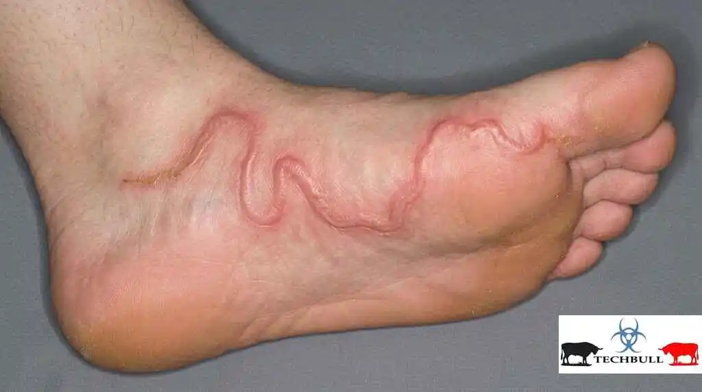
A 34-year-old man presented to an outpatient dermatology clinic ten days after returning from a beach holiday in coastal Brazil, describing a red, thread-like line across the sole of his right foot that appeared to have shifted position overnight. The lesion had begun as a small, itchy papule at a spot where he recalled walking barefoot across damp sand near an area frequented by stray dogs. Over the following days, the papule elongated into a raised, winding track that advanced a few millimeters further each day, and the itching, worse at night, had already cost him two nights of sleep.
This clinical picture — a pruritic eruption that appears to physically travel across the skin over the course of days rather than remaining fixed in one place — is one of the more visually distinctive presentations in clinical dermatology, and also one of the more frequently misdiagnosed, particularly outside endemic regions where clinicians may not immediately recognize it. A migrating, itchy skin lesion is not a single disease; it is a descriptive sign that points toward a specific and fairly narrow set of underlying causes, most of them parasitic, some of them purely inflammatory mimics.
This article establishes a working framework for the migratory pruritic rash: what defines true cutaneous migration as distinct from expansion or recurrence, the anatomy and physiology that make the skin susceptible to larval tunneling, the major named causes and how they are distinguished from one another, a detailed case walkthrough, and current evidence-based approaches to diagnosis and treatment.
Definition and Overview
A migratory pruritic rash refers to a cutaneous lesion — typically a papule, vesicle, wheal, or linear track — that changes location over time, either by physically advancing along the skin surface (as with larval tunneling) or by appearing at new sites in sequence while resolving at old ones (as with certain urticarial and inflammatory conditions). The defining feature that separates it from an ordinary spreading or expanding rash is directional movement: the lesion vacates its original site rather than simply enlarging outward from it.

The prototypical and most commonly encountered cause of a truly migrating cutaneous lesion is cutaneous larva migrans (CLM), also known as creeping eruption, a self-limited zoonotic infection caused by the accidental skin penetration of animal hookworm larvae, most often Ancylostoma braziliense or Ancylostoma caninum. Related but distinct entities — larva currens, loiasis, gnathostomiasis, and several inflammatory mimics such as erythema migrans and dermographic urticaria — share superficial features but differ meaningfully in causative organism, rate of migration, and management, making accurate classification clinically important.
Relevant Anatomy and Mechanism
The skin’s relevance to migratory larval disease lies almost entirely in the architecture of its outermost layers. The epidermis, particularly the stratum corneum and stratum granulosum, is avascular and composed of tightly packed keratinocytes; it offers physical resistance to deeper invasion but is soft enough for a motile larva to tunnel through mechanically and enzymatically. The basement membrane at the dermo-epidermal junction acts as a partial barrier: most zoonotic hookworm species lack the collagenase enzymes required to cross into the dermis efficiently, which is precisely why their larvae become trapped wandering within the epidermis rather than penetrating to deeper tissue.
Human-adapted hookworms, by contrast — Necator americanus and Ancylostoma duodenale — possess the enzymatic machinery to breach the dermis, enter the dermal vasculature, and complete a normal life cycle through the lungs to the intestine. This enzymatic mismatch is the central mechanistic explanation for why animal hookworm larvae in human skin produce a visible, prolonged, intensely itchy surface track rather than a silent, symptom-free tissue migration: the larva is, in effect, an accidental and unsuccessful invader, confined to a compartment it cannot escape, provoking a sustained local immune response as it wanders in search of an exit it will not find.
The itch itself is generated by cutaneous sensory afferents (particularly C-fiber pruriceptors) responding to inflammatory mediators — histamine, tryptase, and eosinophil-derived proteins — released as the host immune system reacts to larval antigens and to physical disruption of keratinocytes along the migration path. Nocturnal worsening of pruritus, a near-universal complaint in these conditions, is attributed to a combination of larval activity that may increase with skin warming under bedding, and the normal circadian reduction in cortisol and rise in core body temperature that lowers the itch threshold overnight.
Pathophysiology: From Skin Penetration to Visible Track
Under normal circumstances, intact stratum corneum functions as an effective barrier against most environmental organisms; disruption typically requires either a breach (abrasion, follicular opening, area of maceration) or an organism specifically adapted to penetrate keratinized tissue using proteolytic secretions. Filariform larvae of zoonotic hookworm species are equipped for exactly this task: they are shed in the feces of infected dogs or cats onto soil or sand, mature to the infective filariform stage in warm, moist, shaded conditions, and actively seek out a host, penetrating bare skin — most often the sole of the foot, buttocks, or hands — within minutes of contact.
Once inside the epidermis, the larva begins to migrate laterally, advancing roughly two to five millimeters per day depending on species and host response. Because it cannot cross the basement membrane into the dermis, it is confined to burrowing through epidermal tissue, which produces the visible, raised, erythematous, serpiginous track that gives the condition its common name. The leading edge of the track — the papule or vesicle marking the larva’s current position — is typically a few millimeters ahead of the visible red trail, since the track itself represents the inflammatory aftermath of tissue the larva has already vacated rather than its present location.
Histologically, the affected epidermis shows spongiosis, a mixed inflammatory infiltrate rich in eosinophils, and, when biopsied precisely at the leading edge, the larva may occasionally be identified within a suprabasal burrow — though biopsy is rarely necessary or advisable given how frequently the larva is missed due to its lag behind the visible track. Peripheral eosinophilia and modestly elevated IgE are common laboratory findings, reflecting a systemic Th2-skewed response to helminth antigen exposure, though these findings are neither sensitive nor specific enough to be diagnostic on their own. Without treatment, the human host represents a dead-end for these zoonotic parasites: larvae cannot complete their life cycle in human tissue and eventually die, usually within two to eight weeks, after which the lesion resolves spontaneously — a fact that shapes counseling even when treatment is offered.
Classification and Key Distinctions
Migratory pruritic skin lesions can be grouped by whether the movement reflects true larval tunneling or a pattern that only mimics migration. The major categories are:
- Cutaneous larva migrans (creeping eruption): Caused most often by Ancylostoma braziliense or A. caninum (cat and dog hookworm). Produces a slowly advancing (2–5 mm/day), intensely itchy, raised, serpiginous track, typically on the feet, buttocks, or hands after barefoot contact with contaminated sand or soil in tropical and subtropical regions.
- Larva currens (Strongyloides stercoralis): A distinct and faster-moving eruption, advancing several centimeters per hour rather than per day, typically around the buttocks, groin, or trunk. Reflects autoinfection by filariform larvae penetrating perianal skin; recurrent episodes over years are characteristic, and hyperinfection is a serious risk in immunosuppressed hosts.
- Loiasis (Loa loa): Produces intermittent, migratory subcutaneous swellings (Calabar swellings) rather than a linear track, along with transient visible migration of the adult worm across the conjunctiva or subcutaneous tissue. Endemic to Central and West Africa; eosinophilia is typically marked.
- Gnathostomiasis: Caused by Gnathostoma species acquired through consumption of undercooked freshwater fish, poultry, or amphibians. Produces migratory, intermittently swelling, tender plaques that can persist for years and, rarely, involve deeper or neurological tissue.
- Erythema migrans (pseudo-migratory mimic): The hallmark early sign of Lyme borreliosis. Technically an expanding, not migrating, annular lesion — it enlarges outward from a fixed tick-bite site rather than relocating — and pruritus is variable to absent, which helps distinguish it from true creeping eruptions despite the overlapping name.
- Dermographic and physical urticaria (pseudo-migratory mimic): Wheals appear, resolve within hours, and recur at new sites, creating an illusion of migration through sequential appearance rather than physical movement of a single lesion. Intensely pruritic but lacks the serpiginous track morphology of larval disease.
Detailed Case Study
Patient Profile
A 34-year-old previously healthy male office worker with no significant past medical history, no regular medications, and no known drug allergies.
History
The patient reported a two-week beach holiday in coastal Brazil, during which he frequently walked barefoot on the sand, including in a shaded area near a beach kiosk where stray dogs commonly rested.
Approximately three days after returning home, he noticed a small, itchy, red bump on the sole of his right foot near the arch. Over the next four days, the bump elongated into a raised, winding, red line that appeared to shift position slightly each morning, extending a few millimeters further than the day before.
Pruritus was severe and consistently worse at night, disrupting sleep. He denied fever, systemic symptoms, joint pain, or lesions elsewhere on the body. He had not sought treatment and had only applied an over-the-counter antihistamine cream with minimal relief.
Examination
A single, raised, erythematous, serpiginous track approximately 4 cm in length was noted on the plantar-medial aspect of the right foot, with a slightly vesicular, tender leading edge and a flatter, drier, hyperpigmented trailing portion.
No surrounding cellulitis, purulent discharge, or lymphangitic streaking was present. No lesions were found elsewhere on examination, including the buttocks, hands, or trunk. Regional lymph nodes were non-palpable and non-tender.
Investigations
The diagnosis was made clinically based on history of barefoot beach exposure and the characteristic morphology; dermoscopy of the leading edge showed a translucent, structureless brownish segment corresponding to the larva’s burrow, supporting the clinical impression.
A complete blood count showed mild peripheral eosinophilia (7% of total white cell count), consistent with but not required for diagnosis. Skin biopsy was not performed, as it was considered unnecessary and unlikely to capture the larva given its typical position ahead of the visible track.
Diagnosis and Management
A diagnosis of cutaneous larva migrans was made on clinical grounds. The patient was treated with a single 200 mcg/kg oral dose of ivermectin, repeated once after one week per standard practice in some protocols, along with an oral second-generation antihistamine for symptomatic pruritus control.
He was counseled on the self-limited but progressive nature of the untreated condition, advised to keep the area clean and avoid scratching to reduce the risk of secondary bacterial infection, and given return precautions for worsening redness, spreading warmth, or purulent discharge suggestive of superinfection. At follow-up ten days later, the track had stopped advancing and pruritus had substantially resolved, with expected post-inflammatory healing over the following weeks.
Associated Conditions, Causes, and Differentials
Beyond the primary parasitic causes of true cutaneous migration, several conditions are relevant either as differentials or as contributing associations clinicians should consider when evaluating a migratory or seemingly migratory pruritic eruption:
- Secondary bacterial infection: Scratching in response to intense pruritus frequently disrupts the skin barrier, permitting staphylococcal or streptococcal superinfection along the track — a common and preventable complication rather than a distinct disease process.
- Scabies (Sarcoptes scabiei): Produces fixed, short, linear burrows rather than progressively advancing tracks, typically in finger webs, wrists, and genital skin, with a characteristic distribution and household clustering that helps distinguish it from CLM.
- Contact and irritant dermatitis: Linear or serpiginous patterns can occur from plant contact (phytophotodermatitis) or irritant exposure, but these lesions are fixed at the site of contact and do not progressively relocate day to day.
- Strongyloides hyperinfection syndrome: In immunocompromised hosts, particularly those on corticosteroids or with HTLV-1 co-infection, larva currens can herald disseminated strongyloidiasis, a potentially fatal complication requiring urgent recognition and treatment.
- Eosinophilic dermatoses: Conditions such as eosinophilic cellulitis (Wells syndrome) can occasionally present with migratory, edematous, itchy plaques and marked tissue eosinophilia, warranting consideration when travel or zoonotic exposure history is absent.
- Insect bite reactions: Sequential, itchy papules from flea, bedbug, or mosquito bites can mimic migration through their linear or clustered distribution pattern, but each individual lesion remains fixed rather than physically advancing.
Comparative Overview of the Three Major Causes
| Feature | Cutaneous Larva Migrans | Larva Currens | Erythema Migrans (Lyme) |
| Causative organism | Animal hookworm larvae (A. braziliense, A. caninum) | Strongyloides stercoralis (autoinfective larvae) | Borrelia burgdorferi (via Ixodes tick bite) |
| Migration rate | 2–5 mm per day | Several centimeters per hour | Not truly migratory; expands outward slowly over days |
| Typical site | Feet, buttocks, hands (sand/soil contact) | Buttocks, groin, trunk (perianal autoinfection) | Site of tick attachment, often trunk or thigh |
| Pruritus | Severe, worse at night | Severe, often described as burning | Variable; often mild or absent |
| Course without treatment | Self-limited, resolves in 2–8 weeks | Chronic, recurrent over years without treatment | Persists and may progress to disseminated Lyme disease |
| First-line treatment | Oral ivermectin or albendazole | Oral ivermectin | Oral doxycycline or amoxicillin |
“The larva that causes a creeping eruption cannot complete its life cycle in human skin — the visible track is the record of a parasite that is, from the outset, already trapped and dying.”
Evidence-Based Management and Practical Guidance
1. Anchor the diagnosis to exposure history
Because the morphology of a migratory pruritic rash can overlap across several conditions, a focused history of recent travel, barefoot contact with sand or soil, freshwater fish consumption, or known tick exposure is often more diagnostically valuable than laboratory testing. In classic presentations, cutaneous larva migrans is diagnosed clinically without the need for biopsy or serology, and treatment should not be delayed pending confirmatory testing that is unlikely to change management.
2. Select antiparasitic therapy based on the likely organism
Oral ivermectin, typically as a single weight-based dose, is regarded as first-line therapy for cutaneous larva migrans in current practice guidance, with oral albendazole as an effective alternative, particularly where ivermectin is unavailable or contraindicated. Topical thiabendazole remains an option for localized, early lesions but is less consistently available and generally less convenient than oral therapy. Larva currens and suspected strongyloidiasis warrant ivermectin with attention to the possibility of underlying chronic infection requiring further evaluation.
3. Manage pruritus and prevent secondary infection
Second-generation oral antihistamines and, where needed, a short course of a mild-to-moderate potency topical corticosteroid can meaningfully reduce discomfort and the urge to scratch while anti parasitic treatment takes effect. Patients should be advised to keep the area clean, avoid vigorous scratching, and monitor for spreading erythema, warmth, or discharge that would suggest secondary bacterial infection requiring antibiotic therapy.
4. Recognize red-flag features that warrant broader workup
Systemic symptoms, marked or persistent eosinophilia, multiple simultaneous lesions, involvement in an immunocompromised patient, or a presentation inconsistent with simple exposure history should prompt consideration of alternative or more serious diagnoses, including loiasis, gnathostomiasis, or Strongyloides hyper infection syndrome, and may justify parasitology referral or further serologic and stool evaluation.
5. Counsel on prevention for future exposure
Because zoonotic hookworm larvae require direct skin contact with contaminated soil or sand to establish infection, footwear on beaches and in areas with known animal fecal contamination, use of a barrier or towel when sitting or lying on sand, and avoidance of walking barefoot in high-risk tropical and subtropical destinations are the most effective preventive measures. Community-level prevention, including deworming programs for domestic and stray animals in endemic coastal areas, addresses the underlying reservoir of infection.
A migratory, pruritic skin rash is a distinctive clinical sign with a relatively narrow and largely recognizable set of causes, dominated in most travel and coastal-exposure contexts by cutaneous larva migrans. Accurate diagnosis rests less on laboratory confirmation than on a careful history of exposure combined with recognition of the characteristic serpiginous, slowly advancing track and its typical distribution on the feet, buttocks, or hands.
Distinguishing true larval migration from faster-moving auto infective conditions like larva currens, from intermittent migratory swellings such as loiasis and gnathostomiasis, and from pseudo-migratory mimics such as erythema migrans and physical urticaria carries direct treatment implications, ranging from a single dose of oral anti parasitic therapy to urgent evaluation for systemic parasitic disease. Red flags that should prompt escalation beyond routine management include systemic symptoms, marked eosinophilia, immunosuppression, multiple or atypical lesions, and any history suggesting freshwater fish consumption or African travel, each of which points toward a less common but more consequential underlying cause.
FAQs
| Question | Answer |
| What does it mean when a skin rash is described as migratory? | It means the visible lesion changes location over time rather than simply enlarging in place, either by a parasite physically tunneling through the skin or by new lesions appearing in sequence as older ones resolve. |
| What is the most common cause of a migratory itchy rash after travel? | Cutaneous larva migrans, caused by animal hookworm larvae penetrating the skin after contact with contaminated sand or soil, is the most common cause in travelers returning from tropical and subtropical coastal regions. |
| How fast does cutaneous larva migrans actually move? | The larval track typically advances two to five millimeters per day, which is visibly noticeable from one day to the next but far slower than the hour-by-hour movement seen in larva currens. |
| Is cutaneous larva migrans contagious from person to person? | No. It is acquired directly from larvae in contaminated soil or sand and is not transmitted from one person to another. |
| Why is the itching worse at night? | Nocturnal worsening reflects a combination of skin warming under bedding, possible increases in larval activity, and the normal circadian drop in cortisol that lowers the overall itch threshold. |
| Can cutaneous larva migrans resolve without treatment? | Yes, because the larva cannot complete its life cycle in human skin and eventually dies, typically within two to eight weeks, but treatment is generally recommended to shorten the course and relieve symptoms sooner. |
| What is the difference between cutaneous larva migrans and larva currens? | Cutaneous larva migrans is caused by animal hookworm larvae and advances slowly over days, while larva currens is caused by Strongyloides stercoralis and can advance several centimeters within a single hour. |
| Where on the body does cutaneous larva migrans typically appear? | It most often affects the soles of the feet, buttocks, and hands, reflecting the body areas most likely to contact contaminated sand or soil. |
| Is a biopsy needed to diagnose a creeping eruption? | Usually not. Diagnosis is typically made clinically based on exposure history and the characteristic appearance, and biopsy often misses the larva because it lies ahead of the visible track. |
| What medications treat cutaneous larva migrans? | Oral ivermectin is generally considered first-line therapy, with oral albendazole as an effective alternative; topical thiabendazole is an option for localized early lesions. |
| Can a migratory rash be a sign of Lyme disease? | Erythema migrans, the early rash of Lyme disease, expands outward from a tick-bite site rather than truly migrating, and pruritus is often mild or absent, which helps distinguish it from larval creeping eruptions. |
| What is loiasis and how does it relate to migratory rashes? | Loiasis is a filarial infection endemic to Central and West Africa that causes intermittent, migratory subcutaneous swellings called Calabar swellings, distinct in distribution and cause from cutaneous larva migrans. |
| Can eating undercooked fish cause a migratory skin rash? | Yes. Gnathostomiasis, acquired through undercooked freshwater fish, poultry, or amphibians, can cause migratory, intermittently swelling, tender skin plaques. |
| Is peripheral eosinophilia expected with these conditions? | Mild eosinophilia is common with cutaneous larva migrans and can be more pronounced in loiasis and gnathostomiasis, though it is not required for diagnosis. |
| How is the diagnosis of cutaneous larva migrans confirmed? | Diagnosis relies primarily on history of relevant exposure and the characteristic serpiginous, advancing track on examination; dermoscopy can support the clinical impression by revealing the larva’s burrow. |
| Are children more susceptible to cutaneous larva migrans? | Children are frequently affected due to more time spent barefoot and playing on beaches or in soil, though the condition can occur at any age with sufficient exposure. |
| Can cutaneous larva migrans be prevented? | Wearing footwear on beaches and soil known to be frequented by animals, and using a barrier when sitting or lying on sand, substantially reduce the risk of larval skin penetration. |
| What happens if cutaneous larva migrans is scratched excessively? | Excessive scratching can disrupt the skin barrier and lead to secondary bacterial infection, which may require additional antibiotic treatment beyond antiparasitic therapy. |
| Is ivermectin safe for treating this condition? | Ivermectin is generally well tolerated and considered safe for cutaneous larva migrans in appropriate candidates, though dosing and suitability should be determined by a healthcare provider based on individual factors. |
| Can pregnant women be treated for cutaneous larva migrans? | Treatment decisions in pregnancy require individualized medical guidance, as some antiparasitic medications are used cautiously or avoided depending on trimester and clinical circumstances. |
| What distinguishes larva currens from ordinary hives? | Larva currens follows a linear or serpiginous track associated with rapid, hour-scale movement near the buttocks or trunk, while urticarial hives are raised wheals that appear and resolve without a true tunneling track. |
| Why is larva currens particularly concerning in immunocompromised patients? | It can signal ongoing autoinfection with Strongyloides stercoralis, which carries a risk of progressing to potentially fatal hyperinfection syndrome in patients who are immunosuppressed. |
| Can a migratory rash indicate a serious internal disease? | In most travel-related cases it reflects a self-limited, treatable skin-confined parasitic infection, but red-flag features such as systemic symptoms or marked eosinophilia should prompt evaluation for more serious underlying causes. |
| How long after exposure do symptoms of cutaneous larva migrans appear? | Symptoms often begin within a few days of skin contact with contaminated sand or soil, though onset can occasionally be delayed by a week or more. |
| Does the rash leave permanent scarring? | Cutaneous larva migrans typically heals without scarring once treated or after spontaneous resolution, barring complications from secondary infection or excessive scratching. |
| Can pets transmit the parasites that cause creeping eruption? | Dogs and cats, particularly strays, can harbor the hookworm species responsible and contaminate soil or sand with larvae through their feces, though the larvae themselves are acquired from the environment rather than direct animal contact. |
| Is repeat infection with cutaneous larva migrans possible? | Yes. Because infection does not confer lasting immunity, repeated exposure to contaminated sand or soil can result in new infections on separate occasions. |
| What should someone do if they notice a migrating itchy line on their skin after travel? | They should seek evaluation from a healthcare provider, mention recent travel and any barefoot exposure to sand or soil, and avoid scratching the area while awaiting assessment. |
| Can over-the-counter creams alone treat cutaneous larva migrans? | Topical antihistamine or steroid creams may ease itching but do not eliminate the larva, so oral antiparasitic therapy is generally required to resolve the underlying infection. |
| Are there long-term effects after successful treatment? | Most patients recover fully without long-term effects after appropriate antiparasitic treatment, with resolution of the track and associated itching within days to a few weeks. |
Medical Disclaimer
This article is intended for general educational purposes only and does not constitute medical advice, diagnosis, or treatment. Readers experiencing a skin rash or other symptoms should consult a qualified healthcare professional for personal evaluation and care. While efforts have been made to ensure accuracy at the time of writing, medical knowledge evolves, and Cloudtechbull.com makes no warranty as to the completeness or current applicability of the information presented.
