
Table of Contents
Deep Vein Thrombosis – The Silent Clot That Catches You Unaware
Understanding the vascular emergency that hides in plain sight — and the everyday habits that can stop it before it starts
It rarely announces itself. There is no dramatic collapse, no gasping first act. A dull ache in the calf after a long flight. A patch of skin that feels warmer than it should. A little swelling you blame on standing too long at work. Deep vein thrombosis, or DVT, is one of the most under-recognized vascular conditions in modern medicine, and that quietness is exactly what makes it dangerous. Somewhere inside a deep vein, usually in the leg, blood that should be flowing freely begins to clump into a soft, sticky clot. Left alone, that clot can grow, can block circulation, and in the worst cases, can break free and travel to the lungs, where it becomes a pulmonary embolism, a genuine medical emergency.
DVT sits at an uncomfortable intersection of common and catastrophic. It is common enough that most people know someone who has had one, whether after surgery, a long-haul flight, or a pregnancy. Yet it is catastrophic enough that, when missed, it ranks among the leading preventable causes of hospital death worldwide. The frustrating part is that DVT is often preventable and, when caught early, highly treatable. The tools to protect yourself are not exotic. They are movement, awareness, hydration, and knowing your own risk profile well enough to act before a clot has the chance to form.
This guide walks through what is actually happening inside the body when a deep vein clot forms, who is quietly at elevated risk without realizing it, the subtle signs that deserve more attention than they usually get, how doctors confirm or rule out a diagnosis, what treatment actually looks like day to day, and the prevention habits worth building into ordinary life. None of this replaces a conversation with a qualified clinician, but understanding the mechanics of DVT is the first real step toward not becoming one of its statistics.

What Deep Vein Thrombosis Actually Is
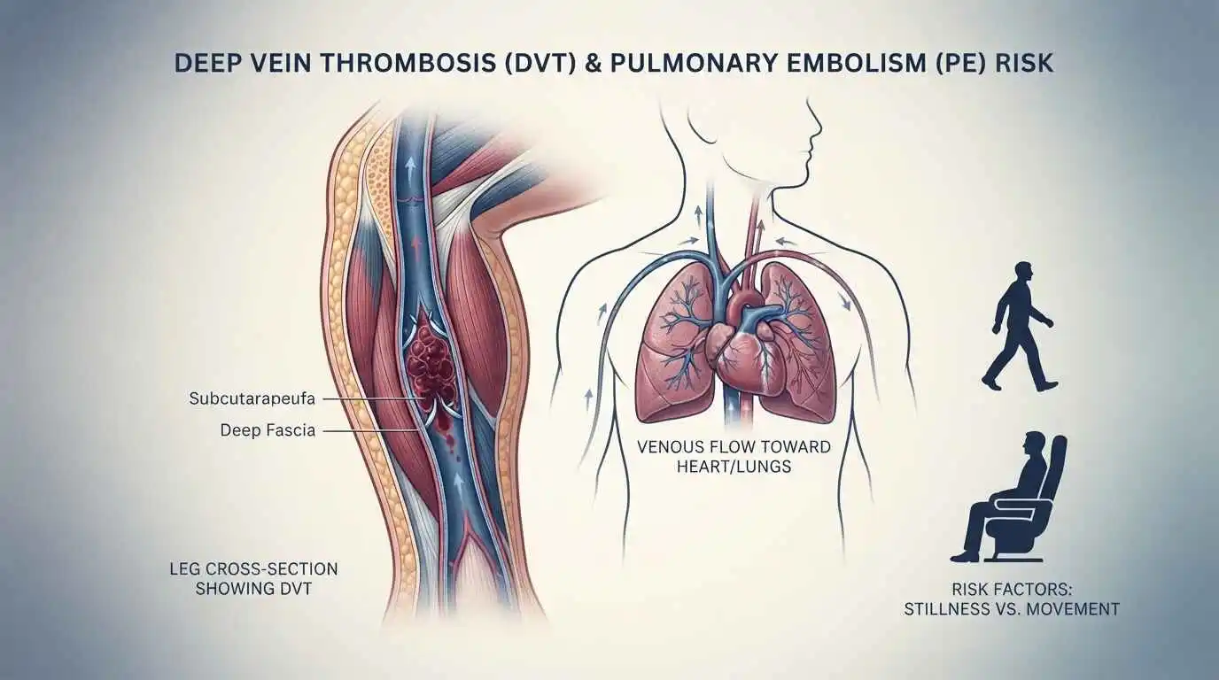
Deep vein thrombosis refers to a blood clot, medically called a thrombus, that forms in one of the body’s deep veins, most commonly in the lower leg, thigh, or pelvis, though it can occasionally develop in the arms. Deep veins are distinct from the superficial veins visible just under the skin. They sit within the muscle tissue and carry the bulk of blood back toward the heart, working against gravity with help from muscle contractions and one-way valves that prevent backflow.
A clot in this system is not automatically a crisis. The body forms small clots routinely as part of normal healing, and the clotting cascade is a vital survival mechanism that stops bleeding after an injury. Trouble begins when that same cascade activates inappropriately inside an intact vein, with nothing to repair, or when a clot grows large enough to obstruct blood flow. As the clot expands, it can partially or completely block the vein, causing blood to back up, pressure to build, and the surrounding tissue to become swollen, tender, and discolored.
The real danger is not usually the clot sitting still in the leg. It is the possibility that a piece of it, or the entire mass, detaches and travels through the venous system, through the right side of the heart, and lodges in the pulmonary arteries supplying the lungs. This is a pulmonary embolism, and depending on its size and location, it can range from mild breathlessness to sudden death within minutes. This is why DVT, even when it feels like a minor leg complaint, is treated with urgency in clinical settings.
The Three Ingredients Behind Every Clot: Virchow’s Triad
Nineteenth-century pathologist Rudolf Virchow described three underlying conditions that, alone or combined, set the stage for abnormal clot formation. Modern vascular medicine still organizes its understanding of DVT around this same framework, generally referred to as Virchow’s Triad.
The first factor is venous stasis, meaning sluggish or stagnant blood flow. Veins rely heavily on muscle movement to push blood back toward the heart. Long periods of immobility, whether from a transatlantic flight, a desk job with minimal movement, bed rest after surgery, or paralysis, all slow this return flow and allow blood to pool in the lower legs. Pooled blood has more time and opportunity to clot.
The second factor is hypercoagulability, a state in which the blood itself is more prone to clotting than usual. This can be inherited, as in genetic clotting disorders like Factor V Leiden or prothrombin gene mutations, or acquired, as seen with pregnancy, certain cancers, hormone therapy, oral contraceptives, dehydration, or systemic inflammation. In each case, the balance between clot-forming and clot-dissolving processes in the blood tips toward formation.
The third factor is endothelial injury, damage to the inner lining of the vein wall. Surgery, trauma, catheter placement, or even chronic inflammation can injure this lining, exposing underlying tissue that actively promotes clot formation as part of the body’s repair response. Most real-world cases of DVT involve more than one of these three factors working together, which is part of why risk tends to cluster around specific life events, like major surgery, cancer treatment, or extended travel, rather than appearing evenly across the general population.
“Roughly half of all deep vein thrombosis cases produce no obvious symptoms — which is exactly why understanding your own risk profile matters as much as recognizing the signs.”
Who Is Quietly at Risk
DVT does not require a dramatic trigger. It often develops from an accumulation of ordinary risk factors that, individually, seem harmless. Recognizing your own risk profile is one of the most practical forms of prevention available.
Age is a meaningful factor, with risk rising steadily after fifty and continuing to climb with each subsequent decade, largely due to changes in vein elasticity and a higher likelihood of coexisting conditions. Recent major surgery, particularly orthopedic procedures involving the hip or knee, dramatically raises short-term risk because of both immobility during recovery and the inflammatory response to tissue trauma. Prolonged immobility more broadly, whether from hospitalization, a cast, long car or plane journeys over four hours, or a sedentary lifestyle, allows blood to stagnate in the calves.
Pregnancy and the postpartum period elevate risk through a combination of hormonal changes, increased clotting factors as a natural safeguard against bleeding during delivery, and physical pressure from the growing uterus on pelvic veins. Hormonal contraceptives and hormone replacement therapy carry a similar, well-documented increase in clotting risk, particularly in combination with smoking. Cancer and its treatments, including chemotherapy and certain catheter placements, are among the strongest risk multipliers, with some tumor types releasing substances that directly promote clotting.
Obesity increases pressure on pelvic and leg veins while often coexisting with reduced mobility. Smoking damages the vascular lining and alters clotting chemistry. A personal or family history of DVT or a known inherited clotting disorder substantially raises the odds of recurrence. Chronic conditions such as heart failure, inflammatory bowel disease, and autoimmune disorders like lupus also contribute meaningfully. None of these factors guarantee a clot will form, but stacking several of them, for instance a smoker recovering from hip surgery on a long flight home, compounds risk sharply, which is exactly why clinicians ask detailed history questions before major procedures.
The Signs Worth Taking Seriously
One of the most dangerous features of DVT is how easily its symptoms are dismissed. Roughly half of all cases produce no obvious symptoms at all, which is part of why screening and risk-based prevention matter so much in high-risk situations like post-surgical recovery.
When symptoms do appear, they typically affect one leg rather than both, since DVT is usually a localized, one-sided event. Swelling is among the most common signs, often noticeable as one calf or thigh appearing visibly larger than the other. Pain or tenderness frequently accompanies the swelling, sometimes described as a cramp or a charley horse that does not resolve with stretching, and classically worsens when the foot is flexed upward, a finding clinicians call Homans’ sign, though it is not reliable enough to use alone for diagnosis.
The skin over the affected area may feel warm to the touch and can take on a reddish or slightly bluish discoloration compared to the surrounding skin. Some people notice the surface veins near the affected area becoming more prominent as blood reroutes around the obstruction. Because these symptoms overlap with far more benign explanations, a pulled muscle, a mild sprain, general fatigue after travel, they are frequently brushed off, particularly by otherwise healthy, active people who do not consider themselves at risk.
The critical distinction is persistence and asymmetry. A cramp that fades within a day is unlikely to be DVT. Swelling and tenderness confined to one leg, that builds rather than improves over twenty-four to forty-eight hours, especially in someone with any of the risk factors above, deserves prompt medical evaluation rather than a wait-and-see approach.
When a Clot Travels: Pulmonary Embolism
The reason DVT commands so much medical respect is its potential to progress into pulmonary embolism, or PE. When a portion of a deep vein clot dislodges, it travels through the venous system, passes through the right chambers of the heart, and becomes lodged in the pulmonary arteries that feed the lungs, blocking blood flow to lung tissue.
Symptoms of PE can develop suddenly and include sharp chest pain that often worsens with deep breathing, shortness of breath that may appear at rest or with minimal exertion, a rapid heart rate, lightheadedness or fainting, and in some cases, coughing up blood-tinged sputum. A large embolism can cause a sudden, severe drop in blood pressure and can be fatal within minutes if the blockage is extensive enough to overwhelm the heart’s ability to pump against it.
Not every embolism is immediately life-threatening; smaller clots may cause milder symptoms that are still frequently missed or misattributed to anxiety, a chest infection, or muscle strain. This is precisely why any sudden onset of chest pain or breathlessness, particularly in someone with known DVT, recent surgery, prolonged travel, or other risk factors, warrants emergency evaluation rather than a wait-and-see approach. Pulmonary embolism remains one of the more common preventable causes of sudden death in hospitalized patients, which is why so much emphasis is placed on clot prevention protocols after surgery.
How Doctors Confirm the Diagnosis
Because DVT symptoms overlap with so many benign conditions, diagnosis relies on a structured combination of clinical judgment and objective testing rather than symptoms alone. Physicians often begin with a standardized risk assessment tool, most commonly the Wells score, which weighs factors like recent immobilization, surgery, swelling, and alternative diagnoses to estimate the probability of DVT before ordering tests.
A D-dimer blood test is frequently used as an initial screening step. D-dimer is a protein fragment released when a clot breaks down, and elevated levels suggest active clotting activity somewhere in the body. The test is highly sensitive, meaning a negative result in a low-risk patient can reliably rule out DVT, but it is not specific: many other conditions, including infection, recent surgery, pregnancy, and even normal aging, can elevate D-dimer levels, so a positive result alone cannot confirm a clot.
Compression ultrasound, specifically duplex ultrasonography, is the primary imaging tool used to confirm DVT. It uses sound waves to visualize blood flow and checks whether a vein compresses normally under gentle pressure from the probe; a vein containing a clot will resist compression. This test is painless, widely available, and does not involve radiation, making it the standard first-line imaging choice.
In more complex or ambiguous cases, physicians may turn to CT venography or MR venography for more detailed imaging, particularly for clots located higher in the pelvis or abdomen that ultrasound cannot easily reach. If a pulmonary embolism is suspected alongside DVT, a CT pulmonary angiogram is typically used to directly visualize the pulmonary arteries. Together, these tools allow clinicians to move efficiently from suspicion to confirmation, which matters enormously given how quickly treatment decisions need to follow a positive diagnosis.
Treating a Clot: What Actually Happens Next
Once DVT is confirmed, the primary treatment goal is preventing the clot from growing, stopping it from breaking loose and reaching the lungs, and reducing the risk of long-term vein damage. The cornerstone of treatment is anticoagulation, commonly known as blood thinning, though the term is slightly misleading since these medications do not actually thin the blood; they interrupt the clotting cascade so that new clot growth slows and the body’s own dissolving mechanisms can gradually break down the existing clot.
Modern treatment typically starts with a direct oral anticoagulant, a category that includes medications working through different points in the clotting pathway, prescribed for a defined course that commonly runs from three months to six months, and sometimes longer for people with ongoing risk factors, recurrent clots, or inherited clotting disorders. In hospitalized or higher-risk patients, injectable anticoagulants may be used initially before transitioning to oral medication. Regular follow-up ensures the dose remains appropriate and monitors for bleeding side effects, which is the primary risk associated with any anticoagulant therapy.
In select severe cases, particularly extensive clots threatening limb viability or large pulmonary emboli causing hemodynamic instability, more aggressive interventions may be used. Thrombolytic therapy involves clot-dissolving medication delivered either systemically or directly at the clot site through a catheter, and can rapidly reduce clot burden but carries a higher bleeding risk, so it is reserved for specific circumstances. Mechanical thrombectomy, physically removing the clot through a catheter-based procedure, is another option in select severe presentations.
For patients who cannot safely take anticoagulants, or who continue to develop clots despite appropriate treatment, an inferior vena cava filter may be placed. This small device sits in the body’s largest vein and is designed to catch clots traveling from the legs before they reach the lungs, without stopping clot formation itself, so it is typically used as a targeted, temporary measure rather than a primary treatment.
Compression stockings are frequently recommended alongside medication, both during acute treatment and afterward, to reduce swelling, support venous return, and lower the risk of long-term complications. Most people manage their DVT treatment as outpatients, attending regular follow-up visits, wearing compression garments as directed, staying appropriately active, and monitoring for any new or worsening symptoms that might signal complications.
Building a Clot-Resistant Routine: Prevention That Works
Prevention strategies split naturally into everyday habits relevant to almost everyone and targeted medical measures reserved for higher-risk situations. On the everyday side, movement remains the single most protective habit available. Calf muscles function as a secondary pump for venous return, so regular contraction, walking, calf raises, ankle circles, or simply standing and stretching every hour during long periods of sitting, keeps blood moving rather than pooling.
During long-haul flights or extended car journeys, aim to stand and walk the aisle every one to two hours where possible, perform seated calf pumps and ankle rotations regularly, and stay well hydrated, since dehydration thickens the blood and raises clot risk. Loose-fitting clothing around the waist and legs supports normal circulation, while compression stockings are a reasonable additional precaution for anyone with existing risk factors making a long journey.
Maintaining a healthy body weight reduces pressure on pelvic and leg veins, while avoiding smoking protects the vascular lining and normal clotting balance. Staying active with regular aerobic exercise supports overall circulatory health well beyond DVT prevention specifically. For people managing chronic conditions that affect mobility, working with a physical therapist to maintain safe movement patterns can meaningfully reduce risk.
On the medical prevention side, anyone undergoing major surgery, particularly orthopedic, cancer-related, or lengthy abdominal procedures, is typically assessed for clot risk beforehand, and many patients receive preventive measures such as early post-operative mobilization, mechanical compression devices worn during and after surgery, or prophylactic low-dose anticoagulation during the highest-risk window of recovery. People with known inherited clotting disorders, a personal history of DVT, or active cancer often work with their physician on a tailored prevention plan that may include longer-term medication, particularly around future high-risk events like surgery, pregnancy, or extended immobility. The overarching principle across all of these measures is the same: keep blood moving, keep the vein lining healthy, and manage the underlying conditions that tip the clotting balance in the wrong direction.
Life After a Clot: Recovery and Long-Term Outlook
Most people who receive prompt, appropriate treatment for DVT recover well, with the clot gradually dissolving or being reabsorbed by the body over weeks to months while anticoagulant therapy prevents new clot growth in the meantime. That said, DVT is not always a one-time event, and understanding the longer arc of recovery matters as much as the initial treatment.
Post-thrombotic syndrome is a chronic complication that develops in a meaningful proportion of people after DVT, caused by lasting damage to the vein valves at the clot site. Symptoms include persistent swelling, aching, heaviness, skin discoloration, and in more severe cases, skin ulcers around the ankle. Wearing compression stockings consistently during and after acute treatment is one of the more effective ways to reduce this risk, along with staying active and elevating the affected leg when resting.
Recurrence is another important consideration. People who have had one DVT carry a higher lifetime risk of a second one, particularly within the first six to twelve months after stopping anticoagulant therapy, which is why the duration of treatment and any decision to extend it indefinitely is individualized based on whether the original clot had a clear, resolved trigger, like surgery, or occurred without any identifiable cause, which generally points toward a higher recurrence risk and a longer treatment horizon.
Emotionally, a DVT diagnosis, especially one accompanied by a pulmonary embolism scare, can understandably leave people anxious about ordinary activities like flying or sitting through a long meeting. Working with a treating physician to understand personal risk level, appropriate precautions, and realistic reassurance around daily activities tends to help far more than blanket avoidance, which is rarely necessary and can itself reduce the very movement that protects against future clots.
Core Lifestyle Changes to Prevent DVT
Medical guidelines consistently emphasize that lifestyle modifications are the first line of defense against venous thromboembolism (VTE) and DVT
- Stay Active and Avoid Prolonged Immobility: Regular movement promotes healthy blood circulation. If you have had surgery, are on bed rest, or are taking a long flight or car ride, make it a habit to move your legs, flex your ankles, or walk for a few minutes every 1–2 hours
- Maintain a Healthy Weight: Excess body weight increases pressure on the veins in your pelvis and legs, significantly elevating your risk of developing a clot
- Stay Hydrated: Drinking plenty of water prevents dehydration, which can cause the blood to become more viscous (thicker) and prone to clotting
- Quit Smoking: Tobacco use damages blood vessel walls and negatively affects blood circulation and clotting mechanisms, making smoking cessation a critical preventive step
- Use Compression Stockings (If Recommended): For individuals with a history of DVT, chronic venous insufficiency, or those undergoing high-risk travel/surgery, graduated compression stockings can help maintain blood flow and prevent pooling in the lower legs
- Elevate Your Legs: When resting, elevating your legs slightly above heart level can assist venous return and reduce swelling
Natural Remedies & Dietary Supports for Vascular Health
While no dietary supplement is as effective in preventing clots as prescription anticoagulants
certain natural foods and compounds can support overall vascular health, reduce inflammation, and promote healthy blood flow as part of a balanced diet.
- Omega-3 Fatty Acids: Found in fatty fish (salmon, mackerel, sardines), flaxseeds, and walnuts, omega-3s have mild antiplatelet effects and reduce systemic inflammation, supporting overall cardiovascular health
- Garlic, Ginger, and Turmeric: These culinary staples contain natural compounds that may help reduce platelet aggregation (clumping) and improve circulation
For example, garlic contains allicin, which has been shown to support healthy blood flow.
- Pycnogenol® (French Maritime Pine Bark Extract): Some clinical studies suggest that taking Pycnogenol before and during long airline flights may help reduce the risk of travel-related DVT by improving venous tone and reducing swelling
- Vitamin E and Antioxidant-Rich Foods: Foods high in vitamin E (such as almonds, spinach, and avocados) and a diet rich in fruits and vegetables help protect blood vessels from oxidative stress
- Horse Chestnut Seed Extract: While primarily studied for chronic venous insufficiency and varicose veins (not active DVT), it may help improve vein tone and reduce leg swelling, indirectly supporting healthy venous return
Critical Cautions Regarding Natural Remedies
If you or your audience are considering natural remedies, it is vital to understand the following risks:
- Dangerous Drug Interactions: Many natural blood-thinning supplements (including high-dose vitamin E, garlic, ginger, ginkgo biloba, and turmeric) can dangerously interact with prescription anticoagulants like warfarin, apixaban, or rivaroxaban, increasing the risk of severe bleeding
- False Sense of Security: Relying solely on natural remedies for DVT prevention in high-risk scenarios (e.g., post-surgery, active cancer, or genetic clotting disorders) is unsafe and medically discouraged
- Vitamin K Consistency: If you are prescribed warfarin, you do not need to avoid vitamin K (found in leafy greens), but you must keep your intake consistent day-to-day, as sudden changes can alter the medication’s effectiveness
When to Seek Immediate Medical Attention
- DVT Signs: Unexplained swelling in one leg, cramping or throbbing pain (often starting in the calf), warm skin, and red or discolored patches on the leg
- PE Signs: Sudden shortness of breath, sharp chest pain that worsens with deep breathing, rapid heart rate, or coughing up blood
The Takeaway
Deep vein thrombosis sits quietly at the edge of everyday life, triggered by ordinary circumstances, a flight, a surgery, a sedentary week, and capable of escalating into a genuine emergency if it goes unnoticed. The encouraging part of this picture is how much control sits within reach. Understanding personal risk factors, staying alert to one-sided leg swelling or pain that does not resolve, moving regularly during long periods of stillness, and seeking prompt evaluation when something feels persistently wrong are all within nearly everyone’s power. None of this is a substitute for professional medical advice, and anyone with symptoms suggestive of DVT or pulmonary embolism should seek medical attention promptly rather than self-diagnose. But armed with a working understanding of how and why these clots form, the silence that makes DVT so dangerous becomes a little easier to break.
| Frequently Asked Questions |
| Q1. What is deep vein thrombosis in simple terms?
It is a blood clot that forms in a deep vein, most often in the leg, which can block normal blood flow and, in serious cases, break loose and travel to the lungs. |
| Q2. What are the earliest warning signs of DVT?
Swelling, aching or cramping pain, warmth, and skin discoloration in one leg are the most common early signs, though about half of all cases show no symptoms at all. |
| Q3. Can DVT occur in the arm instead of the leg?
Yes, though it is far less common. Arm DVT is more often linked to catheters, pacemaker wires, or repetitive strenuous arm activity. |
| Q4. Is DVT the same as a varicose vein problem?
No. Varicose veins involve superficial veins near the skin’s surface, while DVT occurs in the deep venous system within the muscle, and the two are generally unrelated conditions. |
| Q5. How quickly can a DVT turn into a pulmonary embolism?
It can happen within hours to days of clot formation, which is why prompt evaluation of suspicious leg symptoms is strongly encouraged rather than a wait-and-see approach. |
| Q6. Does DVT always cause visible swelling?
No. Swelling is common but not universal, and some clots, particularly smaller or partial ones, produce minimal or no visible swelling. |
| Q7. Can young, healthy people get DVT?
Yes. While risk rises with age, younger people can develop DVT from long flights, hormonal contraceptives, inherited clotting disorders, or after injury and immobilization. |
| Q8. What is Virchow’s Triad?
It is the classic framework describing the three underlying contributors to clot formation: slowed blood flow, increased blood clotting tendency, and injury to the vein’s inner lining. |
| Q9. How is DVT diagnosed?
Diagnosis typically combines a clinical risk assessment, a D-dimer blood test, and a compression ultrasound of the affected limb to visualize the clot directly. |
| Q10. Is the D-dimer test enough to confirm DVT on its own?
No. A negative D-dimer in a low-risk patient can reliably rule out DVT, but a positive result alone cannot confirm it, since many other conditions also raise D-dimer levels. |
| Q11. What does DVT treatment usually involve?
Most cases are treated with a course of oral anticoagulant medication, often for three to six months, sometimes alongside compression stockings and regular follow-up monitoring. |
| Q12. Do blood thinners actually thin the blood?
Not literally. Anticoagulants interrupt the clotting cascade so existing clots can be broken down naturally and new clots are less likely to form, rather than changing blood viscosity. |
| Q13. How long do people typically stay on anticoagulants after a DVT?
It varies, commonly three to six months for a clot with a clear, resolved trigger, and potentially longer or indefinitely for unprovoked clots or recurrent cases. |
| Q14. Are there risks associated with anticoagulant medication?
Yes, primarily an increased risk of bleeding, which is why dosing and duration are individualized and monitored by a treating physician. |
| Q15. What is an IVC filter and when is it used?
It is a small device placed in the body’s largest vein to catch traveling clots before they reach the lungs, generally reserved for patients who cannot safely take anticoagulants. |
| Q16. Can compression stockings prevent DVT?
They can help reduce risk, particularly during long travel or recovery periods, by supporting venous blood flow and reducing pooling in the lower legs. |
| Q17. Is flying a major risk factor for DVT?
Long flights over roughly four hours are associated with increased risk due to prolonged immobility, though the absolute risk for an otherwise healthy traveler remains relatively low. |
| Q18. What can I do on a long flight to reduce DVT risk?
Stand and walk periodically, perform seated calf and ankle exercises, stay hydrated, and avoid tight clothing that restricts circulation in the legs. |
| Q19. Does pregnancy increase the risk of DVT?
Yes, pregnancy and the postpartum period raise clotting risk due to hormonal changes, increased clotting factors, and pressure on pelvic veins from the growing uterus. |
| Q20. Can birth control pills cause DVT?
Hormonal contraceptives are associated with a modestly increased clotting risk, which rises further when combined with smoking or other risk factors. |
| Q21. Is DVT hereditary?
It can be. Inherited clotting disorders such as Factor V Leiden increase susceptibility, and a family history of DVT is considered a meaningful risk factor. |
| Q22. What is post-thrombotic syndrome?
It is a chronic complication that can follow DVT, caused by lasting vein valve damage, leading to persistent swelling, aching, and skin changes in the affected leg. |
| Q23. Can DVT come back after treatment?
Yes, recurrence is possible, particularly within the first year after stopping anticoagulants, which is part of why treatment duration is carefully individualized. |
| Q24. What does a pulmonary embolism feel like?
Common symptoms include sudden chest pain that worsens with breathing, shortness of breath, rapid heart rate, and lightheadedness, and it requires emergency medical attention. |
| Q25. Should I go to the emergency room for suspected DVT?
Persistent one-sided leg swelling or pain warrants prompt medical evaluation, and any accompanying chest pain or breathlessness should be treated as an emergency. |
| Q26. Can exercise help prevent DVT?
Yes, regular movement supports healthy venous return, and activities like walking and calf raises are particularly effective at keeping blood from pooling in the legs. |
| Q27. Does obesity increase DVT risk?
Yes, excess weight increases pressure on pelvic and leg veins and is often associated with reduced mobility, both of which raise clotting risk. |
| Q28. Can surgery trigger DVT even in healthy people?
Yes, surgery combines temporary immobility with tissue trauma and inflammation, which together elevate clotting risk regardless of a person’s baseline health. |
| Q29. Is DVT more common in one leg than both?
Yes, DVT is typically a one-sided, localized event, which is part of why asymmetric swelling or pain is considered a meaningful warning sign. |
| Q30. Can DVT be completely prevented?
Not always, but risk can be substantially reduced through regular movement, hydration, healthy body weight, avoiding smoking, and targeted medical prevention during high-risk periods like surgery or pregnancy. |
Medical Disclaimer: Deep Vein Thrombosis (DVT) is a serious, potentially life-threatening condition. The information below is for educational purposes and should not replace professional medical advice, diagnosis, or treatment. Natural remedies and lifestyle changes are complementary and must never replace prescription anticoagulants (blood thinners) if you are at high risk or have been diagnosed with a clot. The information provided on this website is for general educational and informational purposes only and is not intended as a substitute for professional medical advice, diagnosis, or treatment. Always seek the advice of your physician or other qualified health provider with any questions you may have regarding a medical condition. Never disregard professional medical advice or delay in seeking it because of something you have read on this website.
