
Table of Contents
Peering Inside the Joint: How Arthroscopy Revolutionizes Treatment for Trauma and Sports Injuries
You hear the pop before you feel the pain.
It’s a crisp Sunday morning. You’re playing in your rec league basketball game, you plant your foot to pivot, and suddenly your knee buckles in a direction it absolutely was not designed to go. You hit the hardwood, clutching your leg. In the emergency room a few hours later, the doctor throws around words like “ligament,” “meniscus,” and “cartilage damage.”
Or maybe it’s a rainy Tuesday evening. You’re walking the dog, the pavement is slick, and you take a nasty tumble onto your outstretched hand. Your shoulder immediately screams in protest and refuses to move.
Whether it’s a sudden sports injury on the field or a slip-and-fall at home, acute joint trauma is a terrifying experience. Your mind immediately races to the worst-case scenario: Am I going to be cut open? Am I ever going to get back to normal? Will I have a massive scar and a year of rehab?

Twenty years ago, the answer to severe joint trauma usually involved a large, open incision, days in the hospital, and a long, painful road to recovery. But modern orthopedic surgery has evolved. Today, the gold standard for diagnosing and treating traumatic joint injuries is arthroscopy.
If you or a loved one is facing joint surgery after an accident, you need to understand exactly what this procedure is, how it works, and what your trauma recovery timeline will actually look like. Let’s break down the nuts and bolts of arthroscopic surgery, strip away the medical jargon, and look at how this minimally invasive surgery is getting people back on their feet faster than ever before.
The Intersection of Joint Trauma and Arthroscopic Surgery
To understand why arthroscopy is such a massive leap forward, we first have to understand the nature of joint trauma.
Joints are incredibly complex mechanical structures. Think of your knee or shoulder like the suspension system on a high-performance car. You have the hard framework (the bones), the shock absorbers (the meniscus in the knee, or the labrum in the shoulder), the cables (the ligaments and tendons), and the lubrication (the synovial fluid and articular cartilage).
When you suffer acute joint trauma—whether from a sudden sports injury, a car accident, or a bad fall—that intricate system gets disrupted. A ligament tears. A piece of shock-absorbing cartilage rips and folds over like a bent fender. Sometimes, a chunk of bone gets knocked loose into the joint space, acting like a pebble stuck in a gear.
The Old Way vs. The New Way
In the past, fixing this required open surgery. The surgeon had to make a large incision, cut through healthy muscle and fascia to see the joint, and retract everything out of the way. While this was effective, it caused massive collateral damage. The surgical trauma to the surrounding healthy tissue was often as debilitating as the injury itself. It meant more blood loss, higher infection rates, and weeks of recovery just to heal from the surgery, let alone the original injury.
Enter arthroscopy.
The word literally translates to “looking within the joint.” Instead of opening the whole hood of the car, the mechanic slides a tiny camera through a keyhole to see exactly what is broken, and uses specialized tools through a second keyhole to fix it.
By utilizing arthroscopy, surgeons can visualize, diagnose, and treat joint trauma with unprecedented precision. The trauma to the surrounding healthy tissue is minimized to tiny pokes, rather than long slices. This shift toward minimally invasive surgery is the reason a professional athlete can tear their ACL on a Sunday and be walking on a treadmill by the following weekend.
How Arthroscopy Actually Works: The Tools of the Trade
If you are about to undergo knee arthroscopy or shoulder arthroscopy, it helps to visualize what is actually going to happen in the operating room. Understanding the mechanics of the procedure removes the fear of the unknown.
The Arthroscope: The Surgeon’s Eyes
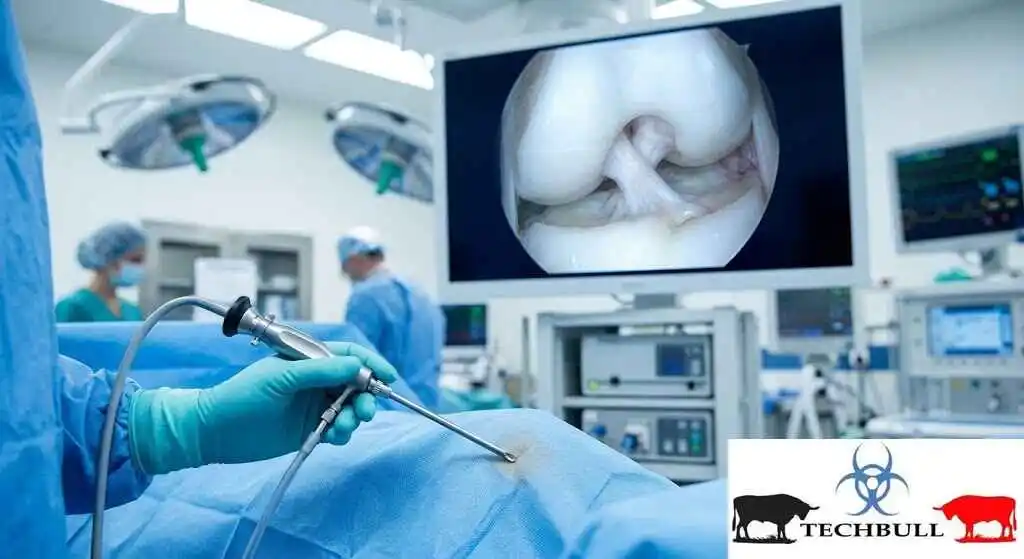
The centerpiece of the entire operation is the arthroscope. This is a rigid, pencil-thin tube containing a high-definition fiber-optic camera and a light source.
During the procedure, the surgeon makes a tiny incision (usually about 5 millimeters long) in the skin over the joint. They insert the arthroscope into this portal. The camera instantly projects a magnified, crystal-clear image of the inside of the joint onto a large high-definition monitor in the operating room.
Because the camera magnifies the area up to 30 times, the surgeon can actually see the damage better than they could with the naked eye during open surgery. They can see the exact texture of the cartilage, the fibers of a torn ligament, and any loose fragments floating in the joint fluid.
The Portals and the Fluid
To make room for the camera and the tools, the joint space has to be expanded. Think of blowing up a balloon; when it’s deflated, the walls are touching. When it’s inflated, there’s a workspace inside.
The surgical team pumps a sterile saline solution into the joint. This distends the joint space, giving the surgeon a clear field of view and washing away any blood or debris from the injury.
The Tools: The Surgeon’s Hands
Once the camera is in place, the surgeon makes one or two additional tiny incisions. These are the “working portals.” Through these holes, they insert specialized, miniature surgical instruments.
These tools are designed to replicate standard surgical instruments but scaled down to fit through a straw.
- Probes: Little hooks used to feel and pull on tissue to test its integrity.
- Graspers: Tiny forceps to grab and remove loose bodies (like bone chips or detached cartilage).
- Shavers: Motorized, rotating blades that suck up and shave away damaged, frayed tissue smoothly.
- Radiofrequency Wands: Tools that use heat to shrink and tighten loose tissue, or to cauterize bleeding vessels.
The Step-by-Step Flow
- Anesthesia: You receive either general anesthesia (you’re asleep) or a regional block (you’re awake but the limb is completely numb and paralyzed).
- Portals: The surgeon marks the anatomy, makes the keyhole incisions, and inserts the camera.
- Diagnostic Sweep: The surgeon systematically moves the camera around the entire joint, assessing the extent of the joint trauma. They document everything. Often, an MRI might miss a small tear, but the arthroscope reveals the true picture.
- The Fix: The surgeon swaps out tools through the working portals to perform the necessary repairs—trimming a torn meniscus, reattaching a torn labrum, or smoothing out cartilage damage.
- Closure: The fluid is drained, the instruments are removed, and the tiny incisions are closed with a single stitch or a small piece of surgical tape. A bulky, sterile bandage is applied.
Common Traumatic Injuries Treated with Arthroscopy
Trauma can strike any joint, but the knees and shoulders take the brunt of the impact in sports and daily accidents. Here is how arthroscopy is used to fix the most common traumatic injuries.
Knee Arthroscopy: The Mechanical Hub
The knee is the most frequently arthroscoped joint in the human body. It takes immense stress, and its reliance on ligaments and cartilage makes it highly susceptible to trauma.
- Meniscus Tears The meniscus is a C-shaped pad of cartilage that acts as a shock absorber between your thigh bone and shin bone. A sudden, forceful twist—like planting your foot and turning on a soccer field—can cause this cartilage to tear.
- The Fix: Using knee arthroscopy, the surgeon evaluates the tear. If the tear is in the “red zone” (the outer third of the meniscus that has blood flow), they will use tiny sutures to stitch it back together so it can heal. If the tear is in the “white zone” (the inner two-thirds, which lacks blood flow and can’t heal), they will use a shaver to carefully trim away only the torn, ragged flap, leaving the healthy cartilage intact.
- ACL Reconstruction The Anterior Cruciate Ligament (ACL) is the primary stabilizer of the knee. When it tears, it rarely heals on its own because it is bathed in joint fluid that prevents clotting.
- The Fix: While the surgeon can’t stitch the ACL back together, they use arthroscopy to reconstruct it. They drill tiny bone tunnels in the thigh and shin, feed a graft (usually a piece of your own hamstring tendon or a donor tendon) through the joint, and secure it in place using specialized buttons or screws.
Shoulder Arthroscopy: The Mobility Master
The shoulder joint is a shallow ball-and-socket, relying heavily on a ring of cartilage called the labrum and a complex web of tendons called the rotator cuff to stay stable.
- Labral Tears (Bankart and SLAP tears) A fall onto an outstretched hand, or a violent dislocation of the shoulder, can rip the labrum right off the bone socket. This causes severe pain, instability, and a feeling that the shoulder will “slip out” during movement.
- The Fix: Through shoulder arthroscopy, the surgeon places tiny plastic or metal anchors into the bone socket. They then pass ultra-strong, non-absorbable sutures through the torn labrum and tie them down to the anchors, effectively re-anchoring the cartilage to the bone.
- Rotator Cuff Repair While some rotator cuff tears are degenerative (from wear and tear over time), acute traumatic tears happen during heavy lifting or sudden falls.
- The Fix: Arthroscopic rotator cuff repair is a highly intricate procedure. The surgeon clears away scar tissue and frayed edges, then places bone anchors into the head of the humerus. They weave sutures through the torn tendon and pull it down to the bone, securing it so the tendon can heal back to its natural attachment point.
Table: Open vs. Arthroscopic Surgery for Joint Trauma
| Feature | Traditional Open Surgery | Arthroscopic Surgery |
| Incision Size | 3 to 6 inches | 2 to 4 tiny incisions (approx. 5mm each) |
| Muscle/Tendon Damage | Requires cutting through healthy tissue | Pushes tissue aside, minimal damage |
| Hospital Stay | 2 to 5 days | Usually outpatient (home same day) |
| Post-Op Pain | High (requires heavy narcotics) | Moderate (managed with NSAIDs/Tylenol) |
| Infection Risk | Higher | Significantly lower |
| Visible Scarring | Large, prominent scar | Barely visible dot scars |
| Time to Return to Sport | 6 to 12 months | 3 to 9 months (depending on injury) |
The Trauma Recovery Timeline: What to Expect After Surgery
One of the biggest misconceptions about minimally invasive surgery is that the recovery is “easy.” The surgical incisions might be tiny, but the internal repair of severe joint trauma is a massive biological event. Your body still has to heal torn ligaments, reattached cartilage, or reconstructed joints.
Trauma recovery is not a straight line; it is a carefully phased progression. If you rush it, you risk tearing the repair. If you are too passive, you will develop permanent stiffness. Here is what the timeline generally looks like.
Phase 1: Acute Healing and Protection (Weeks 0-2)
Immediately after surgery, your joint will be swollen, sore, and wrapped in a bulky dressing. Depending on the trauma, you may be placed in an immobilizer brace or given crutches.
- The Goal: Control inflammation and protect the surgical repair.
- The Action: You will be instructed to elevate the joint above your heart and ice it frequently. You will do gentle, passive range-of-motion exercises to prevent scar tissue from forming, but you will not be bearing weight or doing active lifting.
- Pain Management: Because the surgery was arthroscopic, your pain will peak around day 2 or 3, and then rapidly decline. Most patients transition off prescription painkillers within 5 to 7 days.
Phase 2: Restoring Range of Motion (Weeks 2-6)
This is where the real work begins. The swelling goes down, the incisions heal, and you start physical therapy.
- The Goal: Get your joint moving through a full, normal range of motion without compensation.
- The Action: Your physical therapist will manually stretch the joint and guide you through isolated muscle contractions. If you had knee arthroscopy, you are working on straightening your leg completely and bending it past 90 degrees. If you had shoulder arthroscopy, you are doing pendulum swings and passive lifts.
- The Pitfall: This phase can be frustrating. Progress feels slow, and the joint often feels “tight” or “clicky.” Consistency is key here.
Phase 3: Strength and Neuromuscular Control (Weeks 6-12)
The tissues have healed enough to start taking on real load.
- The Goal: Rebuild the muscle that atrophied (wasted away) after the trauma and surgery, and retrain your brain to use the joint correctly.
- The Action: You transition to active resistance training. Squats, lunges, resistance bands, and light weightlifting. Your therapist will introduce balance and proprioception exercises (like standing on a wobble board) to retrain the tiny stabilizer muscles that protect the joint from future trauma.
Phase 4: Return to Sport/Activity (Months 3-9)
The final phase is about power, agility, and endurance.
- The Goal: Safely return to the activity that caused the injury in the first place.
- The Action: If you are an athlete, you will start a running progression, plyometrics, and sport-specific drills. If you had a cartilage repair, this phase might start later (around month 4 or 5). If you had a simple meniscus trim, you might be back to sports by week 6.
Pro Tip for Recovery: Do not skip your physical therapy “homework.” Going to PT twice a week for an hour is not enough. The 23 hours you spend outside the clinic dictate how well you heal. Set a daily alarm and do your prescribed exercises.
When Arthroscopy Isn’t the Answer (Red Flags and Limitations)
While arthroscopy is a miraculous tool, it is not a magic wand. There are times when joint trauma is too severe, or the patient’s biology is not compatible with a minimally invasive approach. A good surgeon will tell you when arthroscopy is the wrong call.
Severe Osteoarthritis
If an MRI shows that your joint has “bone-on-bone” arthritis, meaning the protective cartilage has completely worn away over the years, arthroscopy is generally not recommended.
Think of it like a car tire. If your tire is bald and completely worn out, using a shaver to smooth out a tiny bubble in the rubber isn’t going to fix the fact that the tire is completely bald. In these cases, an arthroscopic “cleanup” (chondroplasty) provides very short-lived relief. The standard of care for end-stage arthritis is a joint replacement, which requires an open incision to implant metal and plastic components.
Multi-Ligament Trauma and Fractures
If you suffer a high-energy trauma—like a car accident or a fall from a height—you might tear multiple ligaments simultaneously (e.g., ACL, PCL, and MCL in the knee) or suffer fractures that extend into the joint.
While some parts of these complex surgeries might use an arthroscope (like to assist with ACL reconstruction), the surgeon will often need to make open incisions to repair the sheer volume of damaged tissue, stabilize fractures with plates and screws, and ensure the structural integrity of the limb. Trying to fix a knee dislocation entirely through keyhole surgery is risky and time-consuming.
Lack of Post-Op Compliance
A surgeon might elect to use an open approach if they know a patient is unlikely to follow strict physical therapy protocols. Some arthroscopic repairs are highly delicate and rely entirely on the physical therapist to guide the joint through specific ranges of motion safely. If a patient is going to be non-compliant or has neurological issues that prevent them from controlling their muscles, a more robust, open repair with heavier hardware might be necessary to hold the joint together.
FAQ
- What is arthroscopy?
Arthroscopy is a minimally invasive surgical procedure used to visualize, diagnose, and treat problems inside a joint. It uses a tiny camera called an arthroscope inserted through a small incision.
- Is arthroscopy considered major surgery?
It is a surgical procedure, but because it is minimally invasive, it does not carry the same physical toll as major open surgery. Most arthroscopic procedures are done on an outpatient basis, meaning you go home the same day.
- How long does an arthroscopic surgery take?
Depending on the complexity of the joint trauma, the procedure typically takes between 30 minutes to 2 hours. Simple meniscus trims are fast, while ACL reconstructions take longer.
- Will I be awake during the surgery?
Usually, you will be under general anesthesia (fully asleep). However, some procedures can be done with a regional nerve block (where you are awake but the limb is completely numb) combined with mild sedation.
- What are the most common injuries treated by arthroscopy?
The most common include meniscus tears, ACL tears, rotator cuff tears, labral tears in the shoulder, and the removal of loose bone or cartilage fragments resulting from trauma.
- How small are the incisions?
The incisions are incredibly small, usually around 5 millimeters (about a quarter of an inch). You will typically have 2 to 4 of these tiny incisions, often called portals.
- Does arthroscopic surgery hurt?
You will not feel anything during the surgery. Afterward, the joint will be swollen and sore, but because the incisions are tiny and muscles weren’t cut, the pain is significantly less than open surgery. It is usually managed with ice, elevation, and oral painkillers for a few days.
- What is the recovery time for knee arthroscopy?
It depends on what was done. If you had a torn meniscus trimmed, you can walk within a day or two and return to sports in 4-6 weeks. If you had an ACL reconstruction, you will be on crutches for a few weeks and return to sports in 9 to 12 months.
- What is the recovery time for shoulder arthroscopy?
For a simple cleanup, recovery is 4-6 weeks. For a rotator cuff repair or a labral tear repair, you will usually wear a sling for 4-6 weeks, and full recovery takes 4 to 6 months.
- Can I walk immediately after knee arthroscopy?
If you had a simple meniscus trim, yes, you can bear weight immediately, often with crutches for balance for a day or two. If you had an ACL reconstruction or cartilage repair, you will be non-weight-bearing or partial-weight-bearing for several weeks.
- Will I have a scar?
You will have tiny dot scars where the portals were made. These fade significantly over time and are barely noticeable compared to the large scars left by open joint surgery.
- What are the risks of arthroscopy?
While generally safe, risks include infection, blood clots, nerve or blood vessel damage, swelling, and stiffness. The risk of infection is much lower than with open surgery.
- Can a torn meniscus heal on its own without surgery?
If the tear is in the “red zone” (the outer edge with blood supply), it can sometimes heal with rest and physical therapy. Tears in the “white zone” (inner part without blood flow) generally will not heal and require arthroscopic trimming if they are causing pain or mechanical catching.
- Can a torn ACL heal without surgery?
Partial tears might be managed with physical therapy, but a fully torn ACL rarely heals on its own. ACL reconstruction is usually recommended for younger, active patients who want to return to pivoting sports.
- What happens if cartilage damage is left untreated?
Untreated cartilage damage can lead to persistent pain, joint locking or catching, and accelerated osteoarthritis, as the bare bone rubs against other tissues.
- What is a loose body in the joint?
A loose body is a fragment of bone or cartilage that has broken off due to trauma and is floating inside the joint space. It can cause the joint to painfully lock up. Arthroscopy is used to grab and remove these fragments.
- Will I need physical therapy after surgery?
Yes, absolutely. Physical therapy is just as important as the surgery itself. The surgery fixes the anatomy, but PT restores the movement, strength, and proprioception required to use the joint safely.
- Can I drive after arthroscopy?
You cannot drive until you are completely off pain medications and can safely control the pedals (for lower body surgery) or steer without shoulder pain (for upper body surgery). This is usually 1 to 6 weeks, depending on the joint and the repair.
- What is the “clicking” sound in my knee after surgery?
Some clicking or popping is normal in the first few weeks as swelling moves through the joint or as the surgical tools pass over healed tissue. If it is accompanied by sharp pain or locking, contact your surgeon.
- Is arthroscopy covered by insurance?
Yes, if it is deemed medically necessary for treating joint trauma or pathology, it is typically covered by health insurance, though deductibles and copays will apply.
- Can arthroscopy fix arthritis?
No. Arthroscopy cannot cure arthritis. It can be used to clean up loose fragments or smooth frayed cartilage in early-stage arthritis to provide temporary relief, but it will not regrow cartilage or stop the progression of arthritis.
- Why might a surgeon choose open surgery over arthroscopy?
Open surgery might be chosen for severe multi-ligament trauma, joint fractures requiring plates and screws, end-stage arthritis requiring joint replacement, or if the patient’s anatomy makes arthroscopic visualization impossible.
- What is a bone bruise, and can arthroscopy fix it?
A bone bruise is micro-fracturing of the bone due to impact trauma. Arthroscopy cannot fix a bone bruise, as the camera only looks at the surface of the joint. Bone bruises heal with time, rest, and protected weight-bearing.
- How soon after an injury should I have arthroscopy?
It depends. For acute trauma that mechanically locks the joint (like a displaced meniscus tear), surgery may be scheduled within days. For ACL reconstruction, surgeons often wait 3 to 6 weeks for the initial swelling to go down and range of motion to return, which reduces the risk of scar tissue complications.
- What is the difference between an MRI and an arthroscope?
An MRI is a non-invasive imaging test that takes pictures of the inside of the joint. An arthroscope is a surgical tool. Surgeons use MRI to diagnose the problem, but sometimes the MRI misses smaller tears, and the arthroscope reveals the true extent of the damage during surgery.
- Will the hardware used in surgery (screws, anchors) set off metal detectors?
Most surgical hardware is made of titanium or absorbable materials, which generally do not set off standard airport metal detectors.
- Can I tear the same ligament again after arthroscopic repair?
Yes. While the success rate is high, trauma can happen again. Returning to high-risk sports too early, before the graft has fully integrated, is the most common reason for a re-tear.
- What is the most painful part of the recovery?
Usually, the first 48 to 72 hours after surgery, when the joint is swollen and the local anesthetic wears off. After the first week, pain typically drops dramatically and transitions into stiffness and soreness.
- Can I shower after arthroscopy?
You usually need to keep the incisions completely dry for the first 48 hours. After that, you can usually shower with a waterproof dressing, but you cannot submerge the joint in a bath or pool until the incisions are fully healed (usually 2 weeks).
- What is the success rate of arthroscopic surgery for trauma?
It is very high. For common procedures like meniscus trims and ACL reconstructions, success rates are between 85% and 95%, allowing patients to return to their pre-injury level of activity, provided they complete their physical therapy.
Acute joint trauma can feel like a catastrophic end to the activities you love. The pop, the swelling, and the diagnosis of a torn ligament or shredded cartilage can be deeply demoralizing.
But thanks to arthroscopy, a traumatic joint injury is no longer a life sentence of pain and immobility. By utilizing high-definition cameras and micro-instruments, surgeons can meticulously repair the intricate mechanics of your joints without destroying the healthy tissue around them. This minimally invasive surgery means less pain, lower infection risks, and a faster, more aggressive return to rehabilitation.
However, the surgery is only half the battle. The scalpel does the repair, but your dedication to physical therapy dictates your ultimate outcome. Trauma recovery is a marathon that requires patience, consistency, and a willingness to push through the discomfort of rehabilitation.
If you are dealing with a recent injury, don’t wait in the dark. Find an orthopedic specialist who is highly trained in arthroscopic techniques. Get a clear diagnosis, understand the procedure, and start mapping out your recovery timeline. The joint might be broken, but with modern orthopedic surgery, the path back to full strength is clearer than ever before.
⚕️Medical Disclaimer:
The information provided on this website is for general educational and informational purposes only and is not intended as a substitute for professional medical advice, diagnosis, or treatment. Always seek the advice of your physician or other qualified health provider with any questions you may have regarding a medical condition. Never disregard professional medical advice or delay in seeking it because of something you have read on this website.
