
Table of Contents
RUNNING ON EMPTY: The Complete Guide to Every Major Type of Anemia — Causes, Symptoms, Diagnosis & TreatmentBecause Your Blood Tells a Story Your Body Can No Longer Afford to Silence |
Introduction: The Silent Epidemic in Your Blood
Every second, your bone marrow manufactures approximately two million red blood cells — each one a microscopic oxygen-delivery vehicle carrying hemoglobin molecules capable of binding and transporting oxygen from your lungs to every cell in your body. It is one of the most elegant logistics operations in nature. But when that operation is disrupted — when red blood cells are too few, too fragile, too misshapen, or too short-lived — the consequences ripple through every organ system in your body. That disruption has a name: anemia.
Anemia is not a single disease. It is a clinical sign — a measurable deficiency in the blood’s oxygen-carrying capacity — that can arise from dozens of different causes, span genetic inheritance to nutritional neglect, and range in severity from mild fatigue to life-threatening organ failure. The World Health Organization estimates that anemia affects approximately 1.62 billion people globally, making it one of the most prevalent health conditions in the world and the most common nutritional disorder in both developing and developed nations.
India carries a particularly heavy burden. National health surveys consistently reveal that more than half of Indian women of reproductive age are anemic, and nearly two-thirds of children under five suffer from some degree of iron deficiency anemia — numbers that translate directly into reduced productivity, impaired cognitive development, higher maternal mortality, and billions of lost economic outputs each year.
Yet despite its prevalence, anemia is chronically misunderstood. Many people assume it means simply ‘low iron,’ or attribute every fatigue to an iron tablet deficit. The reality is far more nuanced. This guide takes you through the major types of anemia — their mechanisms, causes, symptoms, diagnosis, and treatment — equipping you to understand what your blood is trying to tell you, and what to do about it.

1. Iron Deficiency Anemia — The World’s Most Common Blood Disorder
What It Is and Why It Happens
Iron deficiency anemia (IDA) is the most widespread nutritional disorder on Earth, responsible for roughly half of all anemia cases globally. It occurs when the body’s iron stores fall too low to support normal hemoglobin production, leaving red blood cells small, pale, and poorly equipped to carry oxygen — a pattern pathologists call microcytic, hypochromic anemia.
The causes are multiple and often overlap. Inadequate dietary iron is the most common trigger, particularly in populations dependent on plant-based diets where iron is present in its less bioavailable non-heme form. Chronic blood loss — from heavy menstrual periods, peptic ulcers, colorectal cancer, or regular use of non-steroidal anti-inflammatory drugs (NSAIDs) — is a critical cause that must be excluded in any adult presenting with IDA, particularly men and post-menopausal women for whom dietary causes alone are statistically insufficient. Malabsorption (due to coeliac disease, inflammatory bowel disease, or gastric bypass surgery) and the elevated iron demands of pregnancy round out the most common etiologies.
Symptoms and Consequences
The hallmark symptom is fatigue — a pervasive, bone-deep tiredness that sleep does not relieve. Pallor of the skin, conjunctiva, and nail beds; exertional shortness of breath; heart palpitations; and difficulty concentrating are characteristic. Two unusual signs are worth knowing: pagophagia (craving to eat ice) and koilonychia (spoon-shaped, concave fingernails), both of which, when present, are highly suggestive of IDA. In children, iron deficiency is associated with impaired cognitive development, reduced attention span, and poorer academic performance, even at levels not yet severe enough to cause frank anemia.
Treatment
Oral iron supplementation is the mainstay of treatment — typically ferrous sulfate, ferrous gluconate, or ferrous fumarate, taken between meals with vitamin C to enhance absorption. The underlying cause must simultaneously be identified and addressed; treating IDA without finding the source of iron loss is incomplete medicine. For patients who cannot tolerate oral iron or have severe deficiency, intravenous iron infusions offer a safe and efficient alternative. Dietary counselling to increase consumption of iron-rich foods is essential for long-term prevention.
2. Vitamin B12 and Folate Deficiency Anemia — When DNA Can’t Keep Up
The Megaloblastic Pattern
Vitamin B12 (cobalamin) and folate (vitamin B9) are both essential for the synthesis of DNA, and therefore for the rapid cell division required to produce red blood cells. When either nutrient is deficient, red blood cells cannot divide normally. Instead of producing compact, biconcave discs, the bone marrow generates large, immature, dysfunctional cells called megaloblasts — giving this category its name: megaloblastic anemia.
These enlarged cells have a shortened lifespan and are poorly functional, leading to a macrocytic anemia in which cells are large but ineffective. The neurological consequences of B12 deficiency are distinct from those of folate deficiency and extend well beyond the blood: subacute combined degeneration of the spinal cord — a potentially irreversible neurological syndrome involving damage to the posterior and lateral spinal columns — is a feared complication of prolonged B12 deficiency.
Causes
Vitamin B12 deficiency arises from inadequate dietary intake (strict vegetarians and vegans are at high risk, as B12 is found almost exclusively in animal products), malabsorption due to pernicious anemia (the autoimmune destruction of gastric parietal cells, which produce intrinsic factor — the protein required to absorb B12), gastric surgery, Crohn’s disease, or prolonged use of metformin (which reduces B12 absorption). Folate deficiency typically results from inadequate dietary intake, alcohol excess, pregnancy (when folate demands double), or medications such as methotrexate and trimethoprim that block folate metabolism.
Treatment
B12 deficiency is treated with intramuscular B12 injections (especially in pernicious anemia, where oral absorption is impossible) or, in dietary deficiency, with high-dose oral B12 supplements. Folate deficiency responds rapidly to oral folic acid supplementation. Folic acid supplementation is recommended for all women planning pregnancy to prevent neural tube defects in the developing foetus.
3. Sickle Cell Anemia — A Genetic Code That Changed History
The Molecular Defect
Sickle cell anemia is one of the most studied genetic disorders in human medicine — and one of the most consequential. It is caused by a single point mutation in the gene encoding the beta chain of hemoglobin, substituting the amino acid valine for glutamic acid at position 6. This seemingly small change produces hemoglobin S (HbS), a variant that, under low-oxygen conditions, polymerizes into rigid fibers that distort red blood cells into the characteristic crescent or ‘sickle’ shape that gives the disease its name.
Sickle-shaped cells are rigid, sticky, and short-lived — surviving only 10 to 20 days compared to the normal 120 days for healthy red cells. They clog small blood vessels, triggering painful vascular occlusion crises; they haemolyse (rupture) prematurely, causing chronic hemolytic anemia; and they deprive tissues of oxygen, progressively damaging the spleen, kidneys, brain, lungs, and bones.
Epidemiology and the Malaria Connection
Sickle cell anemia is inherited in an autosomal recessive pattern — meaning that a person must inherit one defective gene from each parent to develop the disease. Carriers (those with one copy of HbS) have the sickle cell trait and are generally healthy, but they carry a significant evolutionary advantage: HbS carriers show substantial protection against severe Plasmodium falciparum malaria. This explains why the sickle cell mutation reached high frequencies in malaria-endemic regions of sub-Saharan Africa, the Middle East, and India’s tribal belt — natural selection favored the carrier state even as it imposed a tragic cost on those inheriting two copies of the gene.
Management
Treatment has advanced significantly. Hydroxyurea, a drug that induces fetal hemoglobin (HbF) production — which does not sickle — is now the cornerstone of long-term management, reducing pain crises, acute chest syndrome, and hospitalizations. Regular blood transfusions, pain management, prophylactic penicillin in children, and pneumococcal vaccination are standard care. Hematopoietic stem cell transplantation offers a potential cure for eligible patients, and gene therapy trials are showing extraordinary early promise.
4. Aplastic Anemia — When the Bone Marrow Falls Silent
Definition and Pathophysiology
Aplastic anemia is a rare but potentially life-threatening condition in which the bone marrow — the spongy tissue within the bones responsible for producing all blood cells — fails catastrophically. Unlike other anemias, aplastic anemia is not just an anemia: it is a pancytopenia, meaning the production of all blood cell lines is impaired simultaneously. Red cell deficiency produces anemia and fatigue. White cell deficiency causes immunosuppression and recurrent infections. Platelet deficiency leads to bleeding and bruising. The combination can be rapidly fatal without treatment.
In the majority of cases (approximately 70–80%), aplastic anemia is idiopathic — meaning no identifiable cause is found — but is believed to be autoimmune in origin, with cytotoxic T-lymphocytes aberrantly attacking and destroying the hematopoietic stem cells of the bone marrow. Known triggers include exposure to certain drugs (chloramphenicol, carbamazepine, NSAIDs), benzene and other industrial chemicals, viral infections (particularly hepatitis viruses, Epstein-Barr virus, and HIV), and radiation exposure.
Diagnosis and Severity
Diagnosis requires a bone marrow biopsy, which reveals a hypocellular marrow — a marrow largely replaced by fat cells, with few or no blood-forming cells. Aplastic anemia is classified as non-severe, severe, or very severe based on the degree of pancytopenia, with treatment urgency escalating accordingly.
Treatment
For young patients with a matched sibling donor, allogeneic hematopoietic stem cell transplantation (bone marrow transplant) is the treatment of choice and offers a cure rate exceeding 80%. For older patients or those without suitable donors, immunosuppressive therapy — typically antithymocyte globulin (ATG) combined with cyclosporine — is the standard approach, achieving response rates of 60–80%. Eltrombopag, a thrombopoietin receptor agonist that stimulates residual marrow cells, has significantly improved outcomes when added to immunosuppression.
5. Hemolytic Anemia — When Red Cells Are Destroyed Too Fast
The Hemolytic Mechanism
Hemolytic anemias form a diverse and complex category united by a single pathophysiological thread: the premature destruction of red blood cells, or hemolysis, at a rate that outpaces the bone marrow’s ability to replace them. Normal red blood cells survive approximately 120 days in the circulation; in hemolytic anemias, this lifespan may be reduced to days or even hours.
Hemolysis can occur within blood vessels (intravascular hemolysis) — releasing free hemoglobin into the plasma, causing hemoglobinuria (dark, cola-colored urine), and producing the characteristic laboratory finding of low haptoglobin — or outside blood vessels in the spleen, liver, and bone marrow (extravascular hemolysis). The clinical presentation depends on whether hemolysis is acute or chronic and whether the bone marrow can compensate sufficiently.
Causes — Intrinsic and Extrinsic
Intrinsic hemolytic anemias arise from defects within the red cell itself. These include enzyme deficiencies such as glucose-6-phosphate dehydrogenase (G6PD) deficiency — the most common human enzyme disorder, affecting over 400 million people globally and causing acute hemolysis triggered by certain foods (fava beans), drugs, or infections — and hereditary spherocytosis, in which membrane protein defects cause spherical, osmotically fragile cells.
Extrinsic hemolytic anemias result from forces outside the red cell. Autoimmune hemolytic anemia (AIHA) occurs when the immune system mistakenly produces antibodies against the patient’s own red cells. Microangiopathic hemolytic anemia (MAHA) arises when red cells are mechanically shredded as they pass through damaged or abnormal small vessels, as occurs in thrombotic thrombocytopenic purpura (TTP), hemolytic uraemic syndrome (HUS), and disseminated intravascular coagulation (DIC). Infection-related hemolysis from malaria — where Plasmodium parasites invade and rupture red cells — is a major cause of hemolytic anemia in tropical regions.
Treatment
Treatment is highly cause-specific. Autoimmune hemolytic anemia responds to corticosteroids, with rituximab, splenectomy, or immunosuppressive agents used in refractory cases. G6PD-deficiency hemolysis is managed by identifying and avoiding triggers. TTP requires urgent plasma exchange. Congenital hemolytic anemias may need chronic transfusion support, splenectomy, or bone marrow transplantation in severe cases.
6. Thalassemia and Anemia of Chronic Disease — Two Underrecognized Burdens
Thalassemia: A Spectrum of Genetic Anemia
Thalassemia is not a single condition but a family of inherited hemoglobin disorders characterized by reduced or absent production of one or more globin chains. Alpha-thalassemia results from deletions or mutations in the alpha-globin genes; beta-thalassemia from mutations in the beta-globin genes. The severity of disease depends on how many gene copies are affected and the nature of the mutations.
At one end of the spectrum, carriers (thalassemia trait or minor) are usually asymptomatic or have only mild microcytic anemia, often misdiagnosed as iron deficiency. At the other extreme, thalassemia major (Cooley’s anemia) presents in infancy with severe, transfusion-dependent anemia, growth failure, bony deformities from marrow expansion, and progressive iron overload from repeated transfusions — a condition that, without proper treatment, is fatal in the first decade of life. India carries the second-largest burden of thalassemia in the world, with approximately 10,000–15,000 thalassemia major children born each year.
Treatment for thalassemia major requires lifelong regular blood transfusions (every 2–4 weeks) combined with iron chelation therapy to prevent iron overload of the heart, liver, and endocrine glands. Hematopoietic stem cell transplantation remains the only established cure; gene therapy approaches targeting the beta-globin gene are in advanced clinical trials and may transform management within the next decade.
Anemia of Chronic Disease
Anemia of chronic disease (ACD), also known as anemia of inflammation, is the second most common cause of anemia globally after IDA, yet it receives far less attention. It develops in the context of prolonged inflammatory states — chronic infections, autoimmune diseases (rheumatoid arthritis, lupus), malignancy, chronic kidney disease, and heart failure.
The pathophysiology is driven by inflammatory cytokines, particularly interleukin-6 (IL-6), which upregulates hepcidin — the liver’s master iron-regulatory hormone. Elevated hepcidin locks iron inside macrophages and liver cells, preventing its release into the circulation for red cell production. Simultaneously, inflammatory cytokines suppress erythropoietin production and shorten red cell survival. The result is a normocytic or mildly microcytic anemia that does not respond to iron supplementation alone and resolves only when the underlying inflammatory condition is controlled.
Treatment of ACD is therefore primarily directed at the underlying disease. In patients with chronic kidney disease-related anemia, erythropoiesis-stimulating agents (ESAs) combined with intravenous iron can effectively raise hemoglobin levels and reduce transfusion dependence. Novel hepcidin antagonists are in clinical development and may offer new therapeutic options.
Conclusion: Your Blood Speaks — Are You Listening?
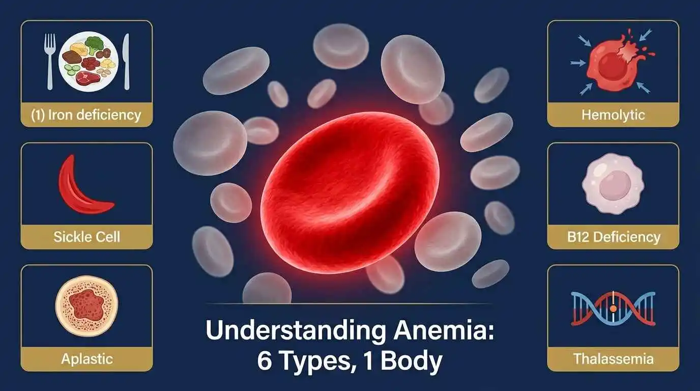
Anemia, in all its forms, is ultimately a message from your body — a signal that somewhere in the intricate chain between bone marrow and bloodstream, oxygen delivery, and cellular renewal, something has broken down. The six major categories explored in this guide — iron deficiency, megaloblastic, sickle cell, aplastic, hemolytic, and thalassemia/chronic disease — each carry a different message, arising from different causes and demanding different responses.
What they share is the capacity to steal vitality quietly, often for months or years before a diagnosis is made. Fatigue becomes ‘normal.’ Breathlessness is attributed to stress. Pallor is dismissed as a complexion. In a country where anemia rates remain alarmingly high across population groups, this silent acceptance carries a heavy collective cost — in productivity, cognitive capacity, maternal health, and child development.
The good news is that most nutritional anemias are preventable and treatable. Genetic forms are increasingly manageable, and the era of gene therapy promises to transform conditions like sickle cell anemia and thalassemia from lifelong burdens into curable diseases. But none of that potential is realized without awareness, accurate diagnosis, and access to care.
Know your hemoglobin. Know your iron. Know your blood. Because running on empty is not a sign of strength — it is a call for help that your body has been making all along.
FAQs — Types of Anemia
| # | Question | Answer |
| 1 | What is anemia? | Anemia is a medical condition in which the blood has a reduced ability to carry oxygen, either due to too few red blood cells, insufficient hemoglobin, or abnormal red blood cell function. |
| 2 | How many types of anemia are there? | There are over 400 types of anemia, broadly categorized into those caused by blood loss, impaired red blood cell production, and increased red blood cell destruction. |
| 3 | What is the most common type of anemia? | Iron deficiency anemia is the most common type worldwide, affecting over 1.2 billion people and disproportionately impacting women of reproductive age and children. |
| 4 | What causes iron deficiency anemia? | It is caused by insufficient dietary iron, poor iron absorption, chronic blood loss (from menstruation, ulcers, or gastrointestinal bleeding), or increased iron demands during pregnancy. |
| 5 | What are the common symptoms of anemia? | Fatigue, weakness, pale or yellowish skin, shortness of breath, dizziness, cold hands and feet, chest pain, and irregular heartbeat are the most common symptoms. |
| 6 | How is anemia diagnosed? | A Complete Blood Count (CBC) is the primary diagnostic test. It measures hemoglobin levels, hematocrit, red blood cell count, and cell size and color to determine the type and severity of anemia. |
| 7 | What hemoglobin level indicates anemia? | The WHO defines anemia as hemoglobin below 13 g/dL in men, below 12 g/dL in non-pregnant women, and below 11 g/dL in pregnant women. |
| 8 | What is pernicious anemia? | Pernicious anemia is an autoimmune condition in which the stomach fails to produce intrinsic factor, a protein needed to absorb vitamin B12, leading to B12 deficiency anemia. |
| 9 | Can anemia be cured? | Many types of anemia are fully treatable and curable (e.g., iron deficiency, B12 deficiency). Others, such as sickle cell and aplastic anemia, are managed rather than cured, though some patients achieve remission through bone marrow transplant. |
| 10 | Is anemia hereditary? | Some forms are genetic, including sickle cell anemia, thalassemia, and hereditary spherocytosis. These are passed from parents to children through specific gene mutations. |
| 11 | What foods help prevent iron deficiency anemia? | Iron-rich foods include red meat, poultry, fish, lentils, beans, tofu, spinach, fortified cereals, and pumpkin seeds. Pairing these with vitamin C enhances iron absorption. |
| 12 | What is megaloblastic anemia? | Megaloblastic anemia occurs when red blood cells are abnormally large and immature due to deficiencies of vitamin B12 or folate, impairing normal DNA synthesis. |
| 13 | What is sickle cell anemia? | Sickle cell anemia is a genetic disorder in which red blood cells become crescent-shaped, sticky, and rigid, blocking blood flow and causing pain crises, organ damage, and chronic anemia. |
| 14 | What is aplastic anemia? | Aplastic anemia is a rare but serious condition in which the bone marrow fails to produce enough blood cells — including red cells, white cells, and platelets — often triggered by autoimmune damage. |
| 15 | What causes hemolytic anemia? | Hemolytic anemia occurs when red blood cells are destroyed faster than the bone marrow can replace them. Causes include autoimmune diseases, infections, medications, blood transfusion reactions, and inherited enzyme defects. |
| 16 | Is anemia dangerous? | Mild anemia may cause only fatigue, but severe or untreated anemia can lead to heart failure, organ damage, pregnancy complications, developmental delays in children, and death in extreme cases. |
| 17 | Can children get anemia? | Yes. Iron deficiency anemia is one of the most common childhood nutritional disorders globally, affecting cognitive development, growth, and immune function. |
| 18 | Is anemia common in India? | India has one of the highest burdens of anemia in the world. The National Family Health Survey (NFHS-5) found that over 57% of women aged 15–49 and 67% of children under 5 are anemic in India. |
| 19 | What is the treatment for anemia? | Treatment depends on the type: iron supplements or dietary changes for iron deficiency; B12 injections or supplements for pernicious anemia; hydroxyurea and blood transfusions for sickle cell; immunosuppressants or bone marrow transplant for aplastic anemia. |
| 20 | Can anemia affect pregnancy? | Yes. Anemia during pregnancy increases the risk of premature birth, low birth weight, maternal mortality, and developmental issues in the baby. Iron supplementation is routinely recommended during pregnancy. |
| 21 | What is thalassemia? | Thalassemia is an inherited blood disorder in which the body produces defective or insufficient hemoglobin. Severe forms (thalassemia major) require regular blood transfusions. |
| 22 | What is anemia of chronic disease? | Also called anemia of inflammation, this type develops alongside chronic conditions such as rheumatoid arthritis, kidney disease, cancer, or HIV, where inflammation impairs iron use and red blood cell production. |
| 23 | Can anemia cause hair loss? | Yes. Iron deficiency anemia is linked to telogen effluvium, a type of hair loss in which hair follicles enter a premature resting phase due to insufficient iron stores. |
| 24 | Does anemia affect mental health? | Chronic anemia, particularly iron and B12 deficiency, is associated with depression, cognitive impairment, memory difficulties, irritability, and brain fog. |
| 25 | Can you have anemia with a normal CBC? | In rare cases, yes — for example, in functional iron deficiency where iron stores are depleted despite normal red cell counts, or in early stages of deficiency anemia. Additional tests such as serum ferritin are then needed. |
| 26 | What is Diamond-Blackfan anemia? | A rare congenital form of pure red cell aplasia, Diamond-Black fan anemia causes the bone marrow to fail to produce red blood cells from birth, requiring lifelong transfusions or steroid therapy. |
| 27 | How does altitude affect anemia? | At high altitudes, the body compensates for lower oxygen by producing more red blood cells. Existing anemia worsens symptoms at altitude as oxygen delivery is further compromised. |
| 28 | Can anemia be prevented? | Many nutritional anemias can be prevented through a balanced diet rich in iron, B12, and folate, food fortification programmes, deworming, and malaria prevention in endemic areas. |
| 29 | What is the link between anemia and fatigue? | Hemoglobin in red blood cells carries oxygen to muscles and organs. When hemoglobin is low, tissues receive less oxygen, causing the characteristic exhaustion and weakness of anemia. |
| 30 | When should I see a doctor about anemia? | Seek medical advice if you experience persistent fatigue, unexplained pallor, breathlessness at rest, heart palpitations, or if blood tests indicate low hemoglobin — do not self-treat with supplements without diagnosis. |
⚕️Medical Disclaimer:
The information provided on this website is for general educational and informational purposes only and is not intended as a substitute for professional medical advice, diagnosis, or treatment. Always seek the advice of your physician or other qualified health provider with any questions you may have regarding a medical condition. Never disregard professional medical advice or delay in seeking it because of something you have read on this website.
