
Table of Contents
Dancing Carotid : Corrigan’s Sign, a Key Indicator of Aortic Regurgitation
As medical aspirants we try learn to interpret the subtle language the human body speaks – a language of signs and symptoms that guide us towards diagnosis. Among the many physical findings we observe, some are particularly striking and hold significant historical and clinical weight. One such sign is the phenomenon known as the “dancing carotid,” or more formally, Corrigan’s sign. This visible, forceful pulsation in the carotid arteries is a classic indicator, often pointing directly towards a significant underlying condition: aortic regurgitation.
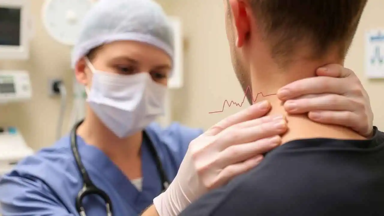
Imagine sitting in a quiet clinic room. A doctor places two fingers gently against the side of your neck, just below the jawline. Instead of feeling a steady, rhythmic thump-thump, the doctor feels a sudden, forceful thrust followed by a rapid, collapsing emptiness—like a water hammer bouncing inside a pipe.
To the untrained eye, nothing looks different. But to a physician, this physical examination finding is a dramatic piece of medical theater. It is known as Corrigan’s Sign, or more evocatively, the “Dancing Carotid” or “Water-Hammer Pulse.”
It is a visceral, tactile clue that hints at a hidden, silent mechanical failure inside the human heart: Aortic Regurgitation (AR).

To understand why an artery in the neck suddenly starts “dancing,” we have to journey inside the chest cavity, explore the architecture of the heart’s most critical exit door, and look at how modern cardiology has evolved to fix this subtle but dangerous defect.
The Man Behind the Sign: Sir Dominic Corrigan
The sign bears the name of Sir Dominic John Corrigan, a distinguished Irish physician. Born in 1802, Corrigan was a keen observer and made significant contributions to the understanding of heart disease, particularly valvular conditions. He meticulously described this characteristic pulse in 1832, noting its association with diseases of the aortic valve. His work highlighted the immense value of careful physical examination, a principle that remains fundamental to medical practice today. When we observe this sign, we are, in a sense, following in the footsteps of this pioneering clinician.
What Exactly is Corrigan’s Sign?
Corrigan’s sign is defined as a visible, bounding, and often palpable pulse in the carotid arteries (located in the neck). While we all have a pulse, Corrigan’s sign is characterized by its exaggerated nature. The carotid arteries appear to visibly pulsate, sometimes with sufficient force to cause a slight bobbing motion of the head or neck (though the head bobbing itself is sometimes referred to as de Musset’s sign, also associated with severe aortic regurgitation).
The term “dancing carotid” is a descriptive and evocative phrase that captures the essence of this sign. It beautifully illustrates the forceful, almost theatrical, movement of the arteries beneath the skin. It’s not just a subtle throb; it’s a prominent, visible dance orchestrated by the powerful dynamics of blood flow.
We typically look for this sign by observing the patient’s neck when they are sitting upright or even lying down with the head slightly elevated. Gentle palpation of the carotid artery can further confirm the bounding, forceful nature of the pulse, often described as feeling like a “water hammer” pulse when felt elsewhere, like the radial artery.
The Root Cause: Aortic Regurgitation
To understand why the carotid arteries perform this “dance,” we must first understand its primary cause: aortic regurgitation (AR).
Aortic regurgitation is a condition where the aortic valve, located between the left ventricle and the aorta, does not close properly after the left ventricle pumps blood out. Instead of forming a tight seal, the valve leaks, allowing a portion of the blood that was just ejected into the aorta to flow back into the left ventricle during diastole (when the heart is relaxing and filling).
This backflow of blood has significant consequences for the heart and the arterial system:
- Volume Overload: The left ventricle receives not only the normal flow of blood from the left atrium but also the blood leaking back from the aorta. This leads to a significant increase in the volume of blood the left ventricle has to handle.
- Ventricular Compensation: The left ventricle compensates over time by dilating (enlarging) and hypertrophying (thickening) its walls. This allows it to hold more blood and pump more forcefully to maintain adequate forward flow despite the backflow.
- Increased Stroke Volume: To compensate for the blood leaking back, the left ventricle ejects a much larger volume of blood with each beat (increased stroke volume).
- Widened Pulse Pressure: This is the key to understanding Corrigan’s sign.
- The increased stroke volume leads to a higher than normal systolic pressure (the pressure in the arteries when the heart contracts).
- The leakage of blood back into the ventricle during diastole, combined with rapid runoff of blood into the peripheral circulation (often exacerbated by peripheral vasodilation in chronic severe AR), causes the pressure in the arteries to drop lower than normal during diastole.
- The difference between the systolic and diastolic pressure is the pulse pressure. In AR, this difference becomes significantly widened.
How Aortic Regurgitation Causes the “Dancing Carotid”
The widened pulse pressure is the direct driver of Corrigan’s sign. Here’s how we understand the mechanism:
- During systole, the massively increased stroke volume is forcefully ejected into the aorta and the large arteries, including the carotids. This causes these elastic vessels to markedly distend. We see and feel this powerful, rapid distension as the peak of the bounding pulse.
- During diastole, the arterial pressure falls rapidly and significantly because of the continuous leak backward through the incompetent aortic valve and the rapid flow away into the periphery. The arterial vessels, having been maximally distended in systole, rapidly recoil as the pressure drops. This rapid collapse is what gives the pulse its characteristic “collapsing” quality, often felt as a sensation like water rushing away under the fingers.
The combination of forceful systolic distension and rapid diastolic collapse creates the large, visible, and bounding pulse wave that we recognize as Corrigan’s sign. The carotid arteries, being large, superficial vessels in the neck, are often where this dramatic pulse is most easily observed.
Clinical Significance of Corrigan’s Sign
Why do we place such importance on spotting a “dancing carotid”? Because, in the context of a cardiac examination, it is a powerful diagnostic clue.
- Strong Indicator of AR: While not exclusively caused by AR (other conditions with high stroke volume and low peripheral resistance can rarely cause it), Corrigan’s sign is highly suggestive of significant aortic regurgitation, particularly when chronic and severe.
- Suggestion of Severity: Generally, the more prominent and visible Corrigan’s sign is, the more severe the underlying aortic regurgitation is likely to be.
- Guides Further Investigation: Finding this sign prompts us to perform a more focused cardiovascular examination and, crucially, to order definitive diagnostic tests like an echocardiogram.
- Part of the Clinical Puzzle: Corrigan’s sign is often present alongside other characteristic physical findings of aortic regurgitation, forming a constellation of signs that paint a clear clinical picture.
Other Classic Signs of Aortic Regurgitation
Corrigan’s sign is just one piece of the clinical mosaic in aortic regurgitation. We are trained to look for several other signs, many of which are also related to the widened pulse pressure and increased stroke volume:
- Water-hammer pulse (or Watson’s pulse): A bounding, rapidly rising and collapsing pulse, typically felt at the wrist (radial artery) upon elevation of the arm. This is essentially the peripheral manifestation of the same phenomenon causing Corrigan’s sign.
- De Musset’s sign: Rhythmical nodding or bobbing of the head with each heartbeat.
- Quincke’s sign: Capillary pulsations visible upon gentle pressure on the nail bed, alternating between flushing and blanching.
- Duroziez’s sign: A systolic murmur heard over the femoral artery upon compression proximal to the stethoscope, and a diastolic murmur heard upon compression distal to the stethoscope.
- Hill’s sign: A significantly higher systolic blood pressure reading in the legs compared to the arms (difference usually >20 mmHg, often much higher in severe AR).
- Traube’s sign (“Pistol shot” femorals): Loud systolic and diastolic sounds heard over the femoral artery.
Observing one or more of these signs, especially Corrigan’s sign, reinforces our suspicion of aortic regurgitation and necessitates further diagnostic workup.
Our Approach to Assessment and Confirmation
When we observe a potential Corrigan’s sign, we perform a thorough cardiovascular examination, including auscultation for the characteristic diastolic murmur of aortic regurgitation heard best at the left sternal border. However, physical examination findings, while invaluable for directing our focus, are not sufficient for definitive diagnosis or assessment of severity.
The gold standard for diagnosing and quantifying aortic regurgitation is echocardiography. This ultrasound imaging of the heart allows us to directly visualize:
- The structure and function of the aortic valve.
- The presence and severity of the regurgitant jet of blood flowing back into the left ventricle.
- The size and function of the left ventricle (assessing for dilation and hypertrophy).
- The pressure gradient across the valve.
Echocardiography provides the detailed information we need to confirm the diagnosis, understand its impact on the heart, determine its severity, and guide management decisions. Other investigations like ECG, chest X-ray, cardiac MRI, and exercise stress tests may also be used depending on the clinical context and plan.
Here’s a simple comparison of typical findings:
| Feature | Normal Carotid Pulse | Corrigan’s Pulse (Severe AR) |
| Visibility | Usually not visibly prominent | Often clearly visible, “dancing” |
| Palpation | Smooth, moderate amplitude | Bounding, rapid rise and fall (“collapsing”) |
| Amplitude | Normal | High amplitude (due to high stroke volume) |
| Pulse Pressure | Typically 30-40 mmHg | Widened (>60 mmHg, often >80 mmHg) |
| Association | Healthy circulation | Suggests significant Aortic Regurgitation |
Management Implications
Identifying Corrigan’s sign fundamentally changes our diagnostic and management pathway. It signals the likely presence of significant aortic regurgitation, a condition that, if severe and symptomatic, often requires surgical intervention (aortic valve replacement or repair) to prevent irreversible damage to the heart muscle and reduce the risk of complications. Even in asymptomatic patients with severe AR, the presence of certain findings (like significant left ventricular dilation) may prompt intervention.
From Suspicions to Certainty: Diagnosing AR
While the “dancing carotid” is a brilliant piece of physical diagnosis, modern medicine relies on imaging to confirm the presence and severity of Aortic Regurgitation.
The Echocardiogram (Echo) is the gold standard. Using sound waves, a cardiologist can watch the aortic valve open and close in real-time. They can measure exactly how much blood is leaking backward (the regurgitant fraction), assess the size of the left ventricle, and determine the exact anatomy of the valve. Is it floppy and stretched out? Is it calcified and rigid?
Once AR is confirmed, the treatment plan depends entirely on one question: Is the heart starting to fail?
Managing the Leak: Treatment Options for Aortic Regurgitation
Unlike some heart conditions that respond well to medication, there is no pill that can fix a leaky valve. However, medications play a crucial supportive role, and surgery offers a definitive cure.
- The “Watch and Wait” Approach (Medical Management)
If the Aortic Regurgitation is mild to moderate, and the left ventricle is not enlarged, doctors usually take a conservative approach. The patient will have an echocardiogram every 1 to 2 years to monitor the heart.
In this phase, if the patient has high blood pressure, doctors will prescribe blood pressure medications (like ACE inhibitors or ARBs). These don’t fix the valve, but they lower the resistance in the arteries, making it physically easier for the heart to pump that extra volume of blood forward, thereby reducing the strain on the left ventricle.
- The Fix: Surgical Aortic Valve Replacement (AVR)
When the leakage becomes severe, the left ventricle begins to enlarge, or the patient develops symptoms (shortness of breath, chest pain, fainting), the valve must be replaced. There is no middle ground.
Historically, this required Open-Heart Surgery (SAVR – Surgical Aortic Valve Replacement). The surgeon makes a large incision down the chest, splits the breastbone, stops the heart using a bypass machine, cuts out the diseased valve, and sews in a new one.
The new valve comes in two main varieties:
- Mechanical Valves: Made of durable materials like titanium or carbon. They last a lifetime, but the patient must take strong blood thinners (Warfarin) for the rest of their life to prevent blood clots from forming on the valve.
- Bioprosthetic (Tissue) Valves: Made from the heart tissue of a cow or pig. They do not require long-term blood thinners, but they typically only last 10 to 15 years before degenerating, meaning a second surgery might be required later in life. (Often chosen for older patients).
- The Minimally Invasive Revolution: TAVR
In recent years, the landscape of Aortic Regurgitation treatment has been dramatically altered by TAVR (Transcatheter Aortic Valve Replacement).
Originally designed for patients too frail to survive open-heart surgery, TAVR is now changing the standard of care. Instead of opening the chest, a cardiologist threads a catheter through an artery in the groin (or occasionally through the ribs or the tip of the heart). A collapsible replacement valve is guided up to the heart and deployed directly inside the old, diseased valve, pushing the old leaflets aside and taking over the job.
Note: While TAVR is now the standard of care for aortic stenosis (a narrowed valve), using TAVR for aortic regurgitation is slightly more complex because the old valve doesn’t have the calcium buildup that helps “anchor” the new TAVR valve in place. However, specialized TAVR devices designed specifically for pure AR are currently in clinical trials and showing immense promise, potentially offering a scar-free, same-day recovery option for the “dancing carotid” patients of the future.
Listening to the Heart’s Whisper
The “Dancing Carotid” is more than just a fascinating relic of 19th-century physical examination; it is a profound reminder of the intricate physics of the human body. A slight mechanical flaw in a valve the size of a quarter can cause arteries in the neck to bounce, heads to bob, and hearts to fail.
Fortunately, we no longer live in Dominic Corrigan’s era. While a physician’s fingertips can still detect the water-hammer pulse, modern echocardiography can see the leak with perfect clarity, and modern surgical techniques—from robotic-assisted open surgery to groundbreaking catheter-based procedures—can repair the broken door.
If you or a loved one have been diagnosed with a heart murmur or Aortic Regurgitation, the most important step is regular monitoring. The heart speaks in whispers before it shouts. By listening closely, and utilizing the incredible treatments available today, we can ensure that the rhythm of the heart remains steady, strong, and securely closed.
Conclusion
Sir Dominic Corrigan’s legacy lives on in the physical examination room. The “dancing carotid,” or Corrigan’s sign, remains a powerful and clinically relevant finding. It serves as a vivid reminder that meticulous observation of the patient’s body can reveal crucial diagnostic information.
“The art of medicine is to be found in observation.” – Sir William Osler
When we observe this bounding, visible pulse, we immediately consider the diagnosis of aortic regurgitation and initiate the appropriate steps for confirmation and management. While modern technology provides us with sophisticated diagnostic tools like echocardiography, signs like Corrigan’s pulse underscore the enduring value of the bedside physical examination. They are threads connecting contemporary medical practice to the foundational principles of clinical observation established by pioneers like Dominic Corrigan, guiding us as we strive to understand and treat the complex conditions affecting the human heart.
FAQs
