
Table of Contents
The Silent Lump That Waited Two Years: Decoding the Subscapular Soft Mass Every Clinician Must Know
When “Nothing” Grows for Two Years
In clinical medicine, the most dangerous presentations are often the quietest ones.
A 45-year-old male walks in — or, more precisely, is eventually persuaded to walk in — with a soft mass beneath his right scapula that has been slowly growing for two years. It doesn’t scream. It doesn’t bleed. It doesn’t wake him in the night with stabbing pain. It just sits there, a polite intruder, announcing itself only as mild discomfort when he lies on his back or rotates his shoulder past a certain angle.
Two years. That is how long this mass was allowed to exist before someone looked at it clinically.
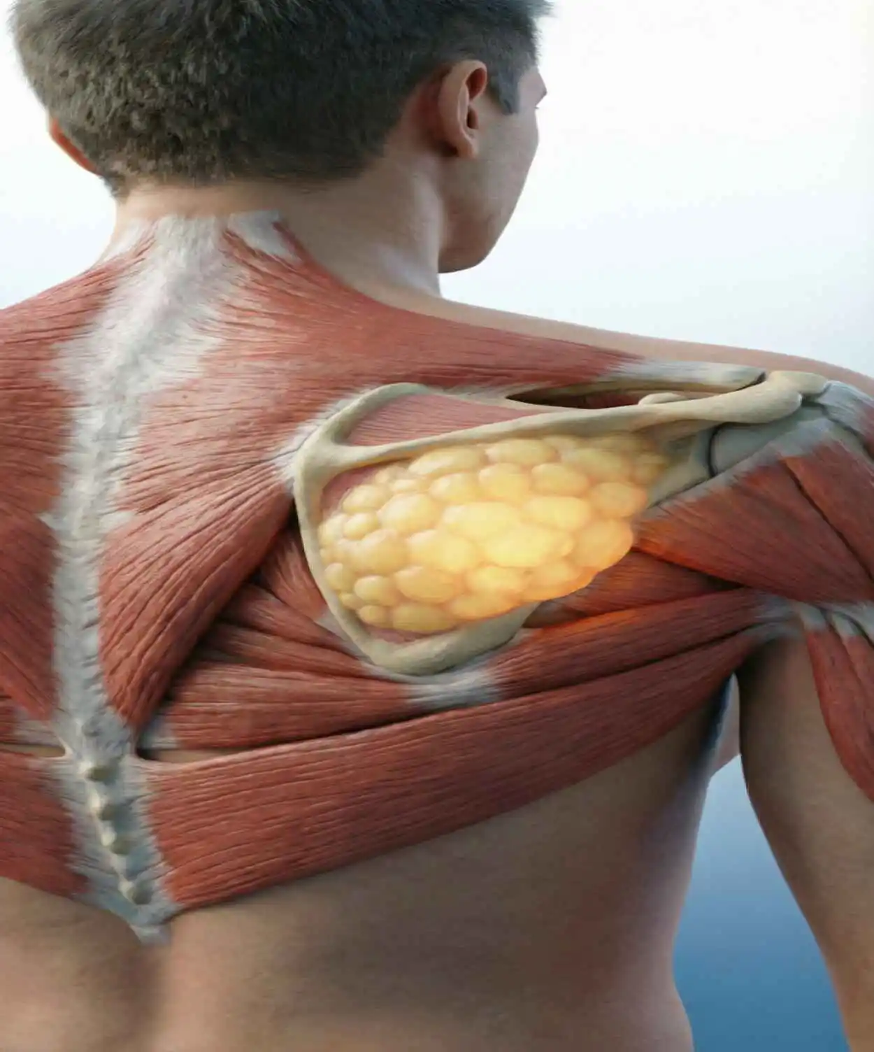
This case, circulated widely in medical education communities, is a masterclass in one of the most instructive tensions in clinical practice: the tension between a presentation that is almost certainly benign and the non-negotiable professional responsibility to rule out what could be catastrophic. The mass in this image — large, soft, clearly delineated, and sitting in the subscapular region with surgical markings already traced upon its surface — tells a story that every clinician, medical student, and surgically curious layperson deserves to understand in full.

Let us decode it, systematically and without flinching.
Reading the Clinical Picture
Before any imaging, any biopsy, or any differential diagnosis algorithm is run, a skilled clinician reads the story of the lesion. And this particular story has several highly informative chapters.
Age and sex: A 45-year-old male. Soft tissue lipomas — the most common benign mesenchymal tumor in adults — peak in incidence between the ages of 40 and 60. Males and females are roughly equally affected, though some studies show a slight male predominance in deeper, intramuscular variants.
Duration: Two years of slow growth. This is one of the most reassuring features in soft tissue mass assessment. Malignant tumors — particularly soft tissue sarcomas — generally grow over weeks to months, not years. A two-year timeline of gradual, continuous, non-accelerating growth strongly favors a benign process.
Character of the mass: Painless. Soft. These two descriptors together are enormously informative. The classic teaching in soft tissue oncology is that a malignant mass tends to be firm to hard in consistency, may be fixed to underlying structures, and often (though not always) becomes painful as it invades adjacent tissues. A freely mobile, soft, compressible mass that has been painless for two years fits the lipoma profile with a high degree of clinical confidence.
Location: Beneath the right scapula — the subscapular region. Lipomas are found throughout the body wherever adipose tissue exists, but the back and posterior trunk are among the most common locations, particularly for larger, deeper-seated variants. Subscapular lipomas can grow to a significant size before becoming symptomatic precisely because the overlying musculature accommodates the mass as it expands.
Symptoms: Mild discomfort when lying supine or with shoulder movement. This symptom pattern is mechanically logical. When the patient lies on his back, the mass is compressed between the scapula and the examination surface, producing pressure-related discomfort. During shoulder movement, the dynamic repositioning of the scapula creates intermittent compression of the lesion. Neither symptom suggests neural invasion, vascular compromise, or aggressive tissue behavior.
Taken together, the clinical history essentially writes its own diagnosis before a single investigation is ordered.
The Differential Diagnosis: What Else Could It Be?
Good clinical practice demands that even a “classic” presentation be systematically considered against its differentials. For a soft subscapular mass in a middle-aged adult, the key entities to consider are:
Lipoma (Most Likely) The straightforward benign adipocytic tumor, composed entirely of mature fat cells enclosed in a thin fibrous capsule. Can be superficial (within the subcutaneous layer) or deep (intramuscular, intermuscular, or subfascial). Deep lipomas, including subscapular variants, tend to be larger at presentation because they are more difficult to detect early. They are the first and dominant diagnosis here.
Lipoma Variants Several lipoma subtypes deserve mention. An angiolipoma contains a vascular component and is typically painful, making it less consistent with this presentation. A fibrolipoma contains fibrous strands and may feel slightly firmer. An infiltrating lipoma or intramuscular lipoma lacks a true capsule, infiltrating between muscle bundles, which can complicate complete surgical excision.
Epidermoid or Inclusion Cyst Epidermoid cysts are common in the back but tend to be more superficial, often with a visible or palpable punctum, and have a firmer, doughy consistency compared to a lipoma’s softer, more fluctuant feel. Unlikely here, given the depth and size.
Elastofibroma Dorsi This is a fascinating and underdiagnosed entity that specifically affects the subscapular region — making it a particularly important differential for this case. Elastofibroma dorsi is a benign reactive fibrous pseudotumor, most commonly found in women over 55, classically located between the inferior angle of the scapula and the chest wall. It presents as a slow-growing, firm mass with a characteristic “winging” of the scapula in some cases. In a 45-year-old male with a soft mass, it is less likely, but the location demands it be on the list.
Desmoid Tumor (Aggressive Fibromatosis): A locally aggressive but non-metastasizing fibroblastic proliferation. Desmoids are firm, not soft, and grow from fascial or musculoskeletal structures. They can occur in the posterior trunk, but the softness and prolonged slow growth make this considerably less likely.
Well-Differentiated Liposarcoma (Atypical Lipomatous Tumor). This is the most critical malignant differential. Well-differentiated liposarcoma is the lowest-grade and most common subtype of liposarcoma, and crucially, it can be clinically and even radiologically indistinguishable from a benign lipoma. It occurs in the retroperitoneum and extremities most commonly, but can arise in the posterior trunk. Features that raise suspicion include large size (>5 cm), deep location, rapid recent change in growth rate, and heterogeneous internal architecture on imaging. This is precisely why no large, deep soft tissue mass should be dismissed as “just a lipoma” without appropriate imaging.
Other Soft Tissue Sarcomas: Myxoid liposarcoma, synovial sarcoma, undifferentiated pleomorphic sarcoma — these are less likely given the clinical features, but remain on the extended differential for any deep soft tissue mass until imaging is reviewed.
The Investigation Pathway
Given the clinical features, the investigation workup follows a logical sequence:
Ultrasound (First-Line Imaging) For superficial or palpably accessible masses, ultrasound is the ideal first investigation. It is fast, radiation-free, inexpensive, and provides immediate information about echogenicity, vascularity (via Doppler), compressibility, and depth. A typical benign lipoma appears as a well-circumscribed, compressible, hyperechoic (or isoechoic) mass with thin internal septations and no significant vascularity. The presence of thick, irregular septations, internal solid components, or pronounced vascularity would raise the suspicion index considerably.
MRI (Definitive Soft Tissue Imaging) For any mass >5 cm, any deep mass, or any lesion with atypical ultrasound features, MRI is the gold standard. Lipomas have a characteristic MRI signature: uniform high signal on T1-weighted sequences (matching subcutaneous fat), uniform low signal on fat-suppressed sequences, and a thin, smooth capsule. Importantly, any internal non-fatty component — nodules, thick septa, areas that do not suppress on fat-sat sequences — demands histological sampling before surgical planning.
Core Needle Biopsy. If imaging suggests any atypical features, image-guided core needle biopsy provides histological confirmation before definitive surgical planning. For lesions with classic lipoma imaging, many surgeons proceed directly to excision. However, for large, deep masses, biopsy-first is increasingly favored to allow appropriate surgical planning — particularly if a sarcoma is in the differential, as it changes the margins required.
CT Chest/Abdomen/Pelvis Reserved for cases where a malignant diagnosis is confirmed or strongly suspected, to stage for metastatic disease.
The Treatment: Surgical Excision
Looking at the image, the surgical planning has already begun. The surgical markings drawn on the skin outline the mass with precision — an elliptical incision pattern designed to allow direct access to the lesion while providing a cosmetically acceptable closure.
For a confirmed or clinically confident benign lipoma, the treatment of choice is surgical excision with the following principles:
Indications for Surgery: Not all lipomas require removal. Small, asymptomatic, superficial lipomas can be monitored clinically. Surgery is indicated when the mass is symptomatic (as in this case), large (>5 cm warrants imaging and surgical discussion), growing, cosmetically concerning, or when the diagnosis is not clinically certain.
Surgical Approach For a subscapular lipoma of this size, the approach involves a direct incision over the mass through skin and subcutaneous tissue, careful dissection through the fascial layers, and identification of the fibrous capsule. The key technical principle is capsule preservation: dissecting along the capsule plane allows the mass to be delivered intact (en bloc), which minimizes local recurrence risk. Piecemeal removal is associated with higher recurrence rates, particularly for intramuscular variants.
If the mass is deep to the scapula or embedded between muscle layers, the dissection requires careful knowledge of regional anatomy — particularly the neurovascular structures of the posterior thoracic wall, including the long thoracic nerve, thoracodorsal nerve, and the subscapular vessels.
Specimen Handling: Even in clinically classic cases, the excised specimen should be sent for histopathological analysis. This is non-negotiable. A mass that looks like a lipoma, feels like a lipoma, and has MRI characteristics of a lipoma can still, rarely, harbor a well-differentiated liposarcoma. Histology closes the diagnostic loop and protects the patient.
Minimally Invasive Alternatives For small superficial lipomas, minimal-incision techniques — where the lipoma is delivered through a small incision using blunt dissection — offer excellent cosmetic outcomes. Power-assisted liposuction has also been described for soft lipomas, though it does not allow specimen retrieval for histology. For a mass of this size and depth, open excision remains the standard.
Recurrence Benign lipomas have low but non-zero recurrence rates. Superficial lipomas, when excised with the capsule intact, rarely recur. Intramuscular lipomas have recurrence rates reported up to 50–60% in some series, particularly if the infiltrative variant is not completely excised.
What Makes This Case Instructive
This is not merely a case about one man’s back lump. It encapsulates several teaching points that are foundational to clinical practice.
Never let patient tolerance determine diagnostic urgency. The patient tolerated this mass for two years. Tolerating a symptom is not the same as the symptom being benign. Many sarcomas — including well-differentiated liposarcoma — present this way. The clinician’s threshold for investigation must not be calibrated by the patient’s pain threshold.
Size and depth are red flags in soft tissue masses. The well-established “5-5 rule” in soft tissue oncology states that any mass greater than 5 cm in diameter or deeper than 5 cm (below the deep fascia) should be investigated with MRI and considered for biopsy, regardless of clinical appearance. The mass in this image is clearly large — likely exceeding 5 cm in diameter.
The subscapular location has a specific differential. Elastofibroma dorsi is a clinical trap specifically in this anatomical region. Any clinician evaluating a subscapular mass must have this entity explicitly in their differential, as its management differs from that of lipoma.
Good surgical markings save operative time. The markings visible in the image show deliberate preoperative planning. Identifying the boundaries of the mass before the patient is anesthetized, positioned, and draped allows for confident, purposeful incision — preventing the disorientation that occurs when a surgeon tries to identify a deep soft mass through surrounding tissue without preoperative surface landmarks.
The Patient Journey Beyond the Operating Room
What happens after the incision is closed matters as much as the operation itself.
Wound care in the posterior back region is complicated by the mechanical demands placed on the skin by shoulder and trunk movement. The wound is under constant dynamic tension, particularly with arm elevation. Careful closure in layers — deep tissue, fascial, subcutaneous, and skin — distributes this tension appropriately. Drains may be placed if the dead space left by removing a large mass is significant, preventing seroma formation.
Recovery depends on the depth and size of the mass. For a lesion of this magnitude, the patient should expect 2–4 weeks of restricted overhead lifting and shoulder activity. Full return to function typically occurs by 6–8 weeks. Follow-up should include wound review at 1–2 weeks and histology result discussion at 2–3 weeks post-operatively.
If histology returns a benign lipoma, reassure, discharge, and counsel on the small recurrence risk. If atypical features or a sarcomatous diagnosis are returned, urgent referral to a specialist soft tissue oncology center is mandatory.
The Broader Lesson: Soft Lumps Are Never Just Soft Lumps
The instinct to reassure — to look at a soft, painless, slow-growing mass and tell a patient “don’t worry, it’s almost certainly nothing” — is not wrong. In the vast majority of cases, it’s right. Most soft masses are lipomas. Most lipomas are benign. Most benign lipomas never cause serious harm.
But clinical medicine is not a game of probabilities without consequences. The rare malignant presentation that is missed because it presented like the common benign one is not a statistical footnote — it is a patient whose outcome was determined, in part, by the clinician’s failure to apply systematic rigor.
The correct approach holds both truths simultaneously: this is almost certainly a lipoma, and I must investigate it appropriately before I can say that with certainty. These are not contradictory positions. They are the simultaneous expression of clinical confidence and clinical humility.
The mass beneath this patient’s right scapula waited two years. The appropriate investigation and treatment should not wait any longer.
Conclusion: Diagnose Deliberately, Operate Carefully, Never Assume
In the clinical image before us, we see more than a soft tissue mass. We see the anatomy of delayed care — a lesion that grew slowly enough to be tolerated, large enough that it now requires a well-planned surgical excision, and located in a region specific enough to demand respect for its differential diagnoses.
The most probable diagnosis is a deep subscapular lipoma. The appropriate management is MRI confirmation, surgical excision with capsule intact, and histopathological analysis of the specimen. The critical professional obligation is to ensure that the comforting clinical picture does not foreclose the disciplined diagnostic process.
This case, simple on its surface, is a reminder that in medicine, the slow and quiet presentations deserve just as much intellectual rigor as the dramatic ones. The lump that doesn’t hurt is not necessarily the lump that doesn’t matter.
Diagnose deliberately. Operate carefully. Never assume.
FAQs
- What is the most likely diagnosis for a painless, slow-growing soft mass beneath the scapula?
The most likely diagnosis is a benign lipoma — specifically a deep or subscapular lipoma — based on the soft consistency, painless nature, two-year slow growth, and characteristic location.
- What is a lipoma?
A lipoma is a benign tumor composed of mature adipose (fat) cells enclosed in a thin fibrous capsule. It is the most common benign soft tissue tumor in adults, typically appearing between the ages of 40 and 60.
- Are lipomas dangerous?
Most lipomas are entirely benign and pose no danger. However, large or deep lipomas should be investigated with imaging to exclude atypical lipomatous tumors or well-differentiated liposarcoma, which can mimic lipomas clinically.
- Why did this patient have discomfort only when lying on his back?
When supine, the mass was compressed between the scapula and the bed surface. During shoulder movement, the repositioning of the scapula intermittently compressed the lesion, both producing mechanical, pressure-related discomfort without indicating malignancy.
- What is elastofibroma dorsi, and why is it relevant here?
Elastofibroma dorsi is a benign fibrous pseudotumor that classically occurs in the subscapular region, making it an important differential for this case. It is more common in older women and typically feels firmer than a lipoma.
- What imaging should be done first for a subscapular soft mass?
Ultrasound is a good first-line investigation for accessible masses. However, for large or deep masses, MRI is the gold standard and provides the most precise characterization of tissue composition and boundaries.
- What does an MRI of a lipoma typically show?
A benign lipoma appears as a uniformly fat-signal mass on T1-weighted MRI (bright, matching subcutaneous fat), with uniform signal dropout on fat-suppressed sequences, and a thin, smooth capsule.
- When should a soft tissue mass be biopsied?
A mass should be biopsied when it is >5 cm, located deep to fascia, shows atypical imaging features, or has changed in growth rate — particularly before surgical planning for potentially malignant lesions.
- What is the “5-5 rule” in soft tissue oncology?
Any mass greater than 5 cm in diameter or deeper than 5 cm below the deep fascia warrants MRI evaluation and potential biopsy, regardless of clinical appearance, due to the increased risk of malignancy.
- Can a lipoma become cancerous?
True benign lipomas essentially never undergo malignant transformation. However, what appears clinically to be a lipoma may occasionally be a well-differentiated liposarcoma from the outset — making histological confirmation after excision essential.
- What is well-differentiated liposarcoma?
It is the lowest-grade subtype of liposarcoma (a malignant fatty tumor) that can be clinically and radiologically indistinguishable from a benign lipoma. It is locally aggressive but rarely metastasizes, and is cured by wide surgical excision.
- What do the surgical markings on the skin represent?
The markings outline the boundaries of the mass on the skin surface, guiding the incision placement and ensuring accurate, deliberate surgical access once the patient is draped and landmarks are harder to identify.
- How is a subscapular lipoma surgically removed?
Through a direct incision over the mass, dissection through fascial layers to the fibrous capsule, and careful en bloc delivery of the intact lipoma. Preserving capsule integrity minimizes recurrence risk.
- Why is en bloc (intact) excision preferred over piecemeal removal?
Intact excision preserves the capsule plane, ensures complete removal, reduces local recurrence risk, and provides a whole specimen for histopathological analysis.
- Is general anesthesia required for lipoma excision?
Small superficial lipomas can be excised under local anesthesia. Large or deep masses like this subscapular lesion typically require general or regional anesthesia for adequate muscle relaxation and patient comfort.
- What are the risks of subscapular lipoma surgery?
Risks include wound infection, seroma formation (fluid collection in the dead space), recurrence, injury to nearby nerves (particularly the long thoracic or thoracodorsal nerve), and — rarely — incomplete excision.
- What happens if a large lipoma is not removed?
Many lipomas remain stable and require no treatment. However, continued growth can cause increasing discomfort, functional restriction, cosmetic concern, and — in rare cases — compression of adjacent neurovascular structures.
- How long is the recovery after subscapular lipoma excision?
Typically, 2–4 weeks of restricted shoulder activity, with full return to function by 6–8 weeks, depending on the size and depth of the excised mass.
- Why must the excised specimen be sent for histopathology?
Even clinically classic lipomas can occasionally harbor atypical histological features. Histopathological analysis is the definitive diagnostic closure — it either confirms benign lipoma or identifies an unexpected pathology requiring further management.
- Can lipomas be dissolved without surgery?
