
Table of Contents
The Silent Storm Within: A Complete Guide to Adrenal Gland Tumors (Pheochromocytoma)
Rare, dramatic, and frequently missed, pheochromocytoma is one of medicine’s most fascinating puzzles. Here is everything you need to understand it.
Imagine a tumor so mercurial it can trigger a catastrophic surge in blood pressure simply from the pressure of a physician’s examining hand. Imagine a condition that mimics anxiety disorders, panic attacks, and hypertension for years before the true culprit is found. Pheochromocytoma is all of this — rare, masterful in its disguise, and life-threatening when missed. This guide illuminates every dimension of this remarkable endocrine tumor.
What Is Pheochromocytoma?
Pheochromocytoma — often abbreviated to “pheo” in clinical settings — is a rare, usually benign tumor that arises from chromaffin cells of the adrenal medulla, the innermost region of the adrenal gland that sits atop each kidney. The name itself derives from the Greek roots for “dusky-colored” (phaios and chroma), referring to the staining characteristics of the tumor cells when exposed to chromium salts in histological preparation.
What makes this tumor so clinically consequential is not merely its existence but its function. Chromaffin cells are the body’s natural factories for catecholamines — specifically epinephrine (adrenaline), norepinephrine (noradrenaline), and occasionally dopamine. These hormones are the biochemical architects of the body’s “fight-or-flight” response. When a pheochromocytoma develops, it produces and secretes these hormones in an unregulated, episodic, and sometimes massive fashion — unleashing physiological storms that can be lethal if unrecognized.

When a similar tumor arises outside the adrenal gland — along the sympathetic ganglia of the abdomen, pelvis, or chest — it is classified as a paraganglioma, a closely related entity sharing many clinical and genetic features with pheochromocytoma. Together, these are often referred to as PPGLs (pheochromocytoma and paraganglioma).
- 2–8 Cases per million people per year
- ~10% of cases are malignant (metastatic)
- 40% of cases have a germline genetic mutation
- 1–0.6% of hypertensive patients have a pheo
The Biology of the Adrenal Medulla
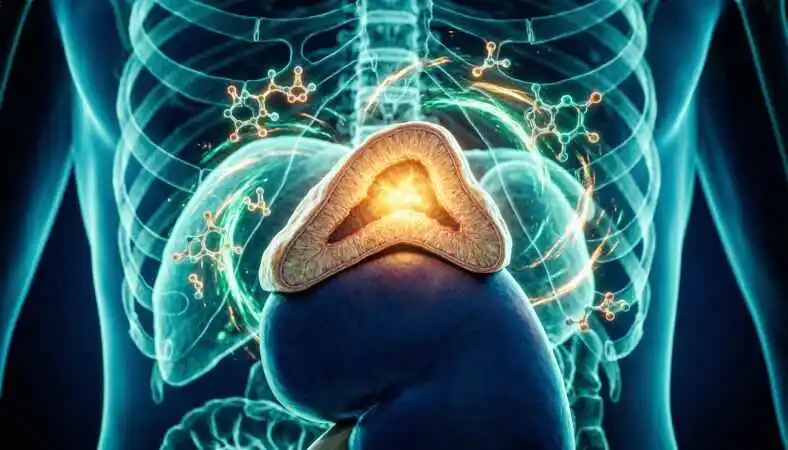
To understand pheochromocytoma, one must first appreciate the organ from which it springs. The adrenal glands are small, pyramidal structures perched atop each kidney. Each gland has two distinct zones: the cortex (outer layer), which produces steroid hormones such as cortisol and aldosterone, and the medulla (inner core), which functions as a specialised part of the sympathetic nervous system.
The medullary chromaffin cells are, in essence, modified postganglionic sympathetic neurons. Rather than releasing neurotransmitters across a synaptic cleft, they secrete catecholamines directly into the bloodstream, acting as endocrine cells. This architecture allows the adrenal medulla to produce rapid, systemic physiological responses — accelerated heart rate, elevated blood pressure, bronchodilation, and mobilisation of glucose — within seconds of a perceived threat.
In pheochromocytoma, this system goes rogue. The tumor cells proliferate abnormally and lose the regulatory feedback mechanisms that normally govern catecholamine release. The result is autonomous, episodic, and sometimes continuous secretion of these potent hormones, with consequences that ripple through virtually every organ system in the body.
Who Gets Pheochromocytoma? Risk Factors and Epidemiology
Pheochromocytoma can affect individuals of any age, though peak incidence occurs between the 30s and 50s. It affects men and women approximately equally. Unlike many cancers, lifestyle factors such as smoking, diet, or alcohol do not appear to increase risk meaningfully. The dominant risk factors are genetic.
Historically described by the “rule of tens” — 10% bilateral, 10% extra-adrenal, 10% malignant, 10% familial — modern genetic analysis has substantially revised this framework. Contemporary research demonstrates that approximately 35–40% of all pheochromocytomas occur in the context of a recognised hereditary syndrome, making genetic testing a cornerstone of management for all newly diagnosed patients.
| Syndrome / Gene | Key Features | Pheo Risk |
| MEN2A / MEN2B (RET) | Medullary thyroid cancer, hyperparathyroidism (2A); marfanoid habitus, neuromas (2B) | 40–50% |
| von Hippel-Lindau (VHL) | Haemangioblastomas, clear-cell renal carcinoma, retinal angiomas | 10–20% |
| Neurofibromatose Typ 1 (NF1) | Café-au-lait spots, neurofibromas, Lisch nodules | 1–5% |
| SDHx mutations (SDHB/C/D/A/AF2) | Hereditary paraganglioma-pheo syndromes; SDHB is strongly associated with malignancy | Variable (up to 70% for SDHD) |
| MAX, TMEM127 | Bilateral adrenal pheo; newer susceptibility genes | Moderate |
The Clinical Presentation: Recognizing the Storm
Pheochromocytoma has earned its reputation as “the great masquerader.” Its symptoms are predominantly driven by catecholamine excess, are often paroxysmal (episodic), and overlap extensively with far more common conditions — particularly essential hypertension, anxiety disorders, and panic attacks.
The classic triad — episodic headache, diaphoresis (profuse sweating), and palpitations — occurs in approximately half of all patients. When these three symptoms occur together in a hypertensive patient, pheochromocytoma should be seriously considered. However, it is important to recognize that this tumor is a consummate chameleon; not all patients present classically, and some tumors are entirely asymptomatic, discovered only incidentally on imaging performed for another reason.
Cardinal Symptoms & Red Flags
- Episodic severe headache
- Profuse sweating episodes
- Palpitations/tachycardia
- Hypertension (sustained or paroxysmal)
- Pallor (not flushing)
- Anxiety, tremor, sense of doom
- Weight loss, heat intolerance
- Hyperglycemia
- Hypertensive crisis during surgery/anesthesia
- Postural hypotension
- Incidentally found an adrenal mass
- Cardiomyopathy (catecholamine-induced)
Attacks — known as “pheo spells” — can last minutes to hours and may be provoked by a variety of stimuli: physical exertion, positional changes (particularly tumors located in the bladder wall, where micturition triggers release), certain foods containing tyramine (aged cheeses, red wine), medications including beta-blockers and some anesthetic agents, and even emotional stress. Between episodes, patients may appear entirely normal, making clinical diagnosis exceptionally challenging.
Diagnosis: Unmasking the Tumor
The diagnostic strategy for suspected pheochromocytoma proceeds in two parallel tracks: biochemical confirmation that excess catecholamines are being produced, followed by anatomical localization of the source.
1 Plasma Free Metanephrines (First-Line Test)
Metanephrines are stable metabolites of catecholamines produced continuously within tumor cells, independent of episodic secretion. Plasma free metanephrine (epinephrine metabolite) and normetanephrine (norepinephrine metabolite) measurements offer sensitivity exceeding 96% for pheochromocytoma, making them the preferred first-line biochemical screens in most international guidelines.
2 24-Hour Urine Catecholamines and Metanephrines
Urine fractionated metanephrines and catecholamines over a 24-hour collection provide complementary biochemical evidence and remain widely used, particularly where plasma assays are unavailable. Specificity is slightly higher than plasma assays, reducing false-positive rates, but sensitivity is marginally lower.
3 CT or MRI of the Abdomen and Pelvis
Once biochemistry is positive, anatomical imaging is the next step. CT with contrast is highly sensitive for adrenal tumors. MRI is preferred when radiation exposure is a concern (children, pregnant women) or when CT is inconclusive. Pheochromocytomas typically appear as large, heterogeneous, vascular masses with areas of hemorrhage or necrosis on imaging. Critical note: Intravenous contrast must only be administered after appropriate alpha-blockade has been established to prevent hypertensive crisis.
4 Functional Nuclear Imaging
For confirming extra-adrenal disease, detecting metastases, or when anatomical imaging is inconclusive, functional nuclear imaging is invaluable. MIBG scintigraphy (metaiodobenzylguanidine labelled with iodine-123) exploits the catecholamine uptake system of chromaffin cells. More recently, PET imaging using 68Ga-DOTATATE (targeting somatostatin receptors) has demonstrated superior sensitivity, particularly for SDHx-related tumors, and is increasingly the preferred functional imaging modality.
5 Genetic Testing
Given that up to 40% of pheochromocytomas are hereditary, germline genetic testing is now recommended for all newly diagnosed patients, regardless of age or family history. A comprehensive gene panel including SDHB, SDHC, SDHD, SDHA, SDHAF2, VHL, RET, NF1, MAX, TMEM127, and others should be offered. Positive results carry profound implications for the patient and their biological relatives.
Pheochromocytoma is frequently found at autopsy in patients who died of seemingly unrelated causes — a sobering reminder that what we do not test for, we cannot find.
— Clinical Endocrinology, International Guidelines
Treatment: Taming the Storm
The definitive treatment for pheochromocytoma is surgical removal of the tumor. However, surgery without adequate medical preparation is one of the most dangerous operations in medicine, carrying the risk of a life-threatening hypertensive crisis on the table. The pre-operative preparation phase is therefore as important as the surgery itself.
Pre-operative Alpha-Blockade
Alpha-adrenergic receptor blockade — typically with phenoxybenzamine (a non-selective, irreversible alpha-blocker) or doxazosin (a selective, reversible alpha-1 blocker) — must be established at least 10–14 days before surgery. This prevents intraoperative hypertensive crises by blocking the peripheral vasoconstriction caused by catecholamine surges during tumor manipulation. A high-sodium diet and generous fluid intake are encouraged concurrently to expand the contracted plasma volume.
Beta-Blockade (After Alpha)
Beta-adrenergic blockade may be added after alpha-blockade is established to control reflex tachycardia. Critical principle: beta-blockers must NEVER be administered before alpha-blockade, as blocking the vasodilatory beta-2 receptors while leaving the vasoconstrictive alpha-1 receptors unopposed can precipitate a catastrophic hypertensive crisis. The sequence — alpha first, beta second — is non-negotiable.
Laparoscopic Adrenalectomy
The gold-standard surgical approach for localized adrenal pheochromocytoma is minimally invasive laparoscopic or retroperitoneoscopic adrenalectomy. Compared with open surgery, the laparoscopic approach yields shorter hospital stays, reduced blood loss, and faster recovery. Posterior retroperitoneoscopic adrenalectomy (PRA) is increasingly preferred at experienced centers for its direct access to the adrenal gland without traversing the peritoneal cavity.
Cortical-Sparing Surgery
For patients with bilateral pheochromocytomas (particularly those with MEN2 or VHL), cortical-sparing (partial) adrenalectomy aims to preserve a rim of functional adrenal cortex, avoiding permanent adrenal insufficiency and lifelong steroid replacement. This technically demanding approach requires experienced surgeons and careful patient selection.
Malignant / Metastatic Disease
For the ~10–17% of pheochromocytomas that are malignant (defined by the presence of metastases in sites where chromaffin tissue does not normally exist), treatment is more complex. Options include 131I-MIBG therapy, peptide receptor radionuclide therapy (PRRT) with 177Lu-DOTATATE, systemic chemotherapy (cyclophosphamide/vincristine/dacarbazine — CVD protocol), and targeted therapies. Prognosis is variable but significantly improved with specialized multidisciplinary management.
Long-Term Surveillance
After successful surgery, all patients require lifelong follow-up. Annual biochemical testing (plasma or urine metanephrines) is recommended for at least ten years, and indefinitely for those with hereditary mutations, bilateral tumors, or extra-adrenal disease. Recurrence risk varies by genotype — SDHB mutation carriers have the highest risk of recurrence and malignancy, requiring particularly vigilant surveillance.
Living with Pheochromocytoma: The Patient Perspective
The journey through a pheochromocytoma diagnosis is rarely straightforward. The average time from symptom onset to diagnosis is reported to be three years or more in many studies — a period often marked by multiple misdiagnoses, dismissed symptoms, and escalating anxiety. Patients who experience episodic attacks frequently report feeling dismissed or told their symptoms are psychosomatic. This diagnostic odyssey takes a significant psychological toll.
Following successful surgery, the majority of patients experience dramatic resolution of their symptoms. Blood pressure normalizes in most cases, episodic attacks cease, and quality of life improves substantially. However, surveillance requirements, the possibility of recurrence, and — for those with hereditary mutations — the knowledge that other family members may be at risk add a layer of ongoing concern that requires supportive, multidisciplinary care.
Patient advocacy organizations and specialist endocrine tumor programs have emerged to address these needs, providing education, peer support, and access to expert care. Awareness remains the critical barrier — for both patients and clinicians — and initiatives to improve recognition of this condition at a primary care level remain urgently needed.
The Future of Pheochromocytoma Research
The landscape of pheochromocytoma research is evolving rapidly. Advances in genomics have already transformed our understanding of disease aetiology, and ongoing work is identifying novel molecular targets for therapy in malignant disease. Liquid biopsy technologies — detecting circulating tumor DNA from blood samples — hold promise for non-invasive monitoring of disease burden and recurrence. Improved functional imaging with next-generation PET tracers is enhancing localization accuracy. And international registries, including the European ENSAT network and the North American NAMSA registry, are pooling data to drive answers that individual centers cannot generate alone. For a disease as rare as pheochromocytoma, international collaboration is not a luxury — it is a necessity.
FAQs
1 What exactly is pheochromocytoma?
Pheochromocytoma is a rare tumor arising from chromaffin cells of the adrenal medulla that autonomously produces and secretes catecholamines — primarily epinephrine and norepinephrine — causing episodic or sustained high blood pressure and a constellation of other cardiovascular and metabolic symptoms.
2 How common is pheochromocytoma?
It is rare, occurring in approximately 2 to 8 people per million per year. It accounts for a secondary cause of hypertension in roughly 0.1–0.6% of hypertensive patients — a small percentage, but given the enormous prevalence of hypertension worldwide, this translates to a substantial number of undiagnosed cases globally.
3 What is the difference between pheochromocytoma and paraganglioma?
Both arise from chromaffin cells and share similar biology and management principles. Pheochromocytoma specifically originates in the adrenal medulla, while paraganglioma arises from extra-adrenal chromaffin tissue — most commonly in the abdomen (retroperitoneal, bladder), but also in the thorax, neck, and skull base. Together, they are classified as PPGLs.
4 Is pheochromocytoma always benign?
No. Approximately 10–17% of pheochromocytomas are malignant, defined by the presence of metastatic disease in sites where chromaffin tissue does not normally exist (bones, liver, lungs, lymph nodes). SDHB mutations are the strongest predictor of malignant behavior. There are no histological criteria that definitively distinguish benign from malignant pheo — metastases are the gold standard of malignancy.
5 What causes a pheochromocytoma to develop?
The precise initiating events are not fully understood. A significant proportion — approximately 35–40% — is caused by germline (inherited) mutations in susceptibility genes including SDHB, SDHC, SDHD, VHL, RET, NF1, MAX, and TMEM127. The remainder are considered sporadic, though somatic (non-inherited) mutations in the same pathways are frequently found in tumor tissue.
6 What does a “pheo spell” feel like?
Patients typically describe a sudden, explosive onset of a severe throbbing headache, drenching sweats, violent palpitations, and a profound sense of anxiety or impending doom. The face is typically pale (not flushed). Blood pressure may spike dramatically — sometimes above 250/150 mmHg. Attacks usually last minutes to an hour and then subside, leaving the patient exhausted.
7 Why is pheochromocytoma so often misdiagnosed?
Because its episodic symptoms — palpitations, anxiety, headaches, sweating, hypertension — closely mimic common conditions such as panic disorder, essential hypertension, hyperthyroidism, and cardiac arrhythmias. Attacks may be infrequent, and between episodes, patients appear entirely normal. Most clinicians encounter only a handful of cases in a career, reducing the index of suspicion.
8 What is the best test to screen for pheochromocytoma?
Current international guidelines — including those from the Endocrine Society and European Society of Endocrinology — recommend plasma-free metanephrines as the first-line biochemical test due to their high sensitivity (exceeding 96%). Urine fractionated metanephrines are a widely used alternative, particularly where plasma testing is not available.
9 Can medications cause false positive results?
Yes. Tricyclic antidepressants, levodopa, sympathomimetics, buspirone, some antipsychotics, and stimulant medications can all elevate metanephrine levels and produce false positive results. Acetaminophen can interfere with some laboratory assays. A careful medication history and, where possible, drug washout periods before testing are important for accurate interpretation.
10 Why must alpha-blockade come before beta-blockade?
Beta-blockers block the vasodilatory beta-2 adrenergic receptors. If given without prior alpha-blockade, the vasoconstrictive alpha-1 receptors remain unopposed, leading to severe peripheral vasoconstriction and potentially catastrophic hypertensive crisis. Alpha-blockade must always be established first to neutralise this risk before any beta-adrenergic antagonism is introduced.
11 Is surgery always necessary?
For localized, respectable pheochromocytoma, surgical removal is the definitive and strongly preferred treatment. Medical management alone with alpha-blockade controls symptoms but does not eliminate the tumor or its long-term risks. For patients with inoperable or metastatic disease, non-surgical therapies including MIBG radionuclide therapy, PRRT, and chemotherapy become the mainstays of management.
12 What happens if a pheochromocytoma is left untreated?
Untreated pheochromocytoma carries a substantial risk of life-threatening hypertensive crisis, stroke, myocardial infarction, aortic dissection, and catecholamine-induced cardiomyopathy. It is among the causes of preventable sudden death. Even asymptomatic tumors can discharge massive catecholamine loads unpredictably — during physical exertion, minor trauma, or anesthesia induction.
13 Can pheochromocytoma cause heart failure?
Yes. Prolonged or massive catecholamine excess can cause catecholamine-induced cardiomyopathy — a potentially reversible form of dilated cardiomyopathy characterized by ventricular dysfunction, reduced ejection fraction, and heart failure symptoms. This complication can be the presenting feature of pheochromocytoma and may resolve dramatically after tumor removal.
14 Can pheochromocytoma affect blood sugar levels?
Yes. Catecholamines stimulate glycogenolysis and gluconeogenesis in the liver and inhibit insulin secretion by the pancreas. This results in elevated blood glucose (hyperglycemia) during attacks and may produce a diabetes-like state. In some patients, apparent new-onset diabetes resolves completely after tumor resection.
15 What is an incidentaloma, and how does it relate to pheochromocytoma?
An adrenal incidentaloma is an adrenal mass discovered on imaging performed for an unrelated reason. They are found in approximately 3–5% of abdominal CT scans. All adrenal incidentalomas require biochemical evaluation to exclude pheochromocytoma before any invasive procedure — including biopsy — is performed, as unrecognized pheo and needle biopsy can trigger a hypertensive crisis.
16 Is pheochromocytoma hereditary?
In approximately 35–40% of cases, yes. Genetic mutations in susceptibility genes are transmitted in an autosomal dominant pattern in most syndromes. A positive genetic result in a patient has direct implications for first-degree relatives, who should be offered cascade genetic testing and appropriate surveillance if a mutation is identified.
17 Can pheochromocytoma occur in children?
Yes, though it is uncommon. In children and adolescents, pheochromocytoma has a higher rate of hereditary association — up to 70% in some pediatric series — is more likely to be bilateral or extra-adrenal, and requires specialist pediatric endocrine management. Any child presenting with hypertension should have pheochromocytoma considered in the differential diagnosis.
18 Can pheochromocytoma occur during pregnancy?
Yes, and this is one of the most clinically dangerous scenarios. Pheochromocytoma in pregnancy carries significant maternal and foetal mortality if unrecognized. It can mimic pre-eclampsia. If diagnosed in the first or early second trimester, laparoscopic resection after alpha-blockade is feasible. After 24 weeks, elective caesarean section with simultaneous or subsequent adrenalectomy is generally preferred.
19 What imaging technique is most reliable for locating the tumor?
CT of the abdomen and pelvis is the initial anatomical imaging modality of choice due to its high sensitivity for adrenal masses. MRI is preferred in specific situations. For functional confirmation and extra-adrenal/metastatic disease, 68Ga-DOTATATE PET/CT has emerged as the most sensitive functional imaging technique and is now recommended preferentially over MIBG scintigraphy by many expert centers.
20 What foods or substances should be avoided with pheochromocytoma?
