
Table of Contents
Anuric Patients: A Complete Guide to Causes, Diagnosis, and Modern Management
Let me paint a picture you’ve probably seen before.
It’s 3 AM in the ICU. A post-surgical patient who was stable six hours ago now has an empty catheter bag. Not low output. Empty. The nurse flags it. The resident orders a bladder scan. Nothing. They push fluids. Nothing. They check the chart and realize the last urine output documented was over four hours ago.
That moment—when the bag is empty and the clock is ticking—is where anuria enters the conversation. And it’s a conversation that too few clinicians are fully prepared for, because anuria sits at the crossroads of so many different disease processes that it can feel overwhelming.
This guide is meant to change that. Whether you’re a nephrology fellow, an ICU nurse, a medical student, or someone trying to understand a family member’s condition, this is everything I know about anuric patients—organized, practical, and written the way I’d explain it to a colleague.

What Is Anuria? Clearing Up the Confusion
Anuria is defined as urine output of less than 100 milliliters per day in adults. Some textbooks use a cutoff of 50 mL per day, and a few define it as essentially no urine output at all. The exact number matters less than the clinical reality: the kidneys have stopped producing urine in any meaningful way.
The word itself comes from Greek—*an-* meaning “without” and ouron meaning “urine.” Simple origin. Complex implications.
Here’s where people get confused: anuria is not the same as oliguria. Oliguria is reduced urine output, typically defined as less than 400 mL per day. Anuria is the extreme end of that spectrum—the point where the kidneys aren’t just struggling, they’ve effectively stopped.
That distinction isn’t academic. It changes your differential diagnosis, your urgency, and your management. A patient with oliguria might still be making 200 mL a day. Their kidneys are compromised but not silent. An anuric patient is in a different category of trouble.
The Three Doors: Prerenal, Intrinsic Renal, and Postrenal
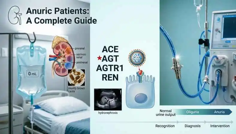
Every case of anuria walks through one of three doors. This framework is the backbone of nephrology, and it’s especially critical in anuria because the causes—and the interventions—diverge so dramatically.
Prerenal Anuria: The Kidney Itself Isn’t the Problem
In prerenal anuria, the kidney tissue is intact. The problem is that blood isn’t getting to it in adequate volume or pressure. The filters work fine. The pipes bringing blood to the filters are the issue.
Think of a water purification plant. The filters are perfectly functional. But the reservoir is empty, or the pipes are crimped. The plant can’t produce clean water not because the filters are broken, but because nothing is flowing in.
Common prerenal drivers include:
- Severe hypovolemia from hemorrhage, dehydration, or massive gastrointestinal losses
- Shock—cardiogenic, septic, or hemorrhagic
- Heart failure where forward flow is insufficient to perfuse the kidneys
- Renovascular obstruction from thrombi or emboli
The clinical tell is often the response to fluid resuscitation. If you restore volume and the kidneys wake up, you’ve found your culprit. That reversibility is what makes prerenal disease so important to identify early. Prolonged ischemia can convert a functional prerenal state into structural damage—and that’s when the story gets worse.
Intrinsic Renal Anuria: The Kidney Tissue Is Damaged
Here, the problem is inside the kidney. The filters themselves are compromised.
Acute tubular necrosis (ATN) is the heavyweight champion of intrinsic renal anuria. It happens after prolonged ischemia, sepsis, or exposure to nephrotoxins like aminoglycosides, contrast dye, or certain chemotherapy agents. The tubular epithelial cells take a hit and stop functioning properly.
But ATN isn’t the only player. The differential for intrinsic anuria includes:
- Acute glomerulonephritis, especially rapidly progressive forms with hematuria, proteinuria, and red cell casts
- Acute interstitial nephritis, often drug-induced (proton pump inhibitors, NSAIDs, beta-lactams), sometimes with fever, rash, and eosinophilia
- Thrombotic microangiopathy (HUS/TTP), where microthrombi clog the kidney’s small vessels
- Rhabdomyolysis, where muscle breakdown products overwhelm the tubules
- Hepatorenal syndrome in patients with advanced liver disease
Intrinsic renal anuria is generally less reversible in the short term than prerenal disease. The damage is structural. Recovery depends on the kidney’s ability to regenerate tubular cells—which it can do, but it takes time. Weeks, sometimes months.
Postrenal Anuria: Something Is Blocking the Drain
This is the plumbing problem. Urine is being produced, but it can’t get out.
Postrenal causes of anuria are almost always obstructive. In adults, the most common culprits are:
- Bilateral ureteral obstruction from stones, tumors, or retroperitoneal fibrosis
- Prostate enlargement or prostate cancer causing bladder outlet obstruction
- Clot retention after urologic surgery or trauma
- Ureteral obstruction in a solitary kidney
Here’s the critical thing about postrenal anuria: it’s often reversible, and time matters enormously. A ureteral obstruction relieved within days often allows full recovery of renal function. Weeks of obstruction? The damage may become permanent.
The clinical clue is often hydronephrosis on ultrasound, or a palpable bladder in the case of outlet obstruction. Bladder scan first, then renal ultrasound. That sequence will save you time and catch the easy diagnoses before you go down rabbit holes.
The Genetic Connection: When Anuria Is Written in the DNA
Some anuria isn’t caused by trauma, infection, or drugs. It’s caused by a faulty gene.
Renal tubular dysgenesis (RTD) is a rare but devastating condition where the proximal tubules of the kidney fail to develop properly in utero. The result is anuria from birth—or even before birth, manifesting as severe oligohydramnios because fetal urine is the main component of amniotic fluid.
RTD is caused by mutations in genes that regulate the renin-angiotensin system:
- ACE (angiotensin-converting enzyme)
- AGT (angiotensinogen)
- AGTR1 (angiotensin II receptor type 1)
- REN (renin)
These genes work together in a cascade that produces angiotensin II, a protein that constricts blood vessels and regulates blood pressure. When the cascade is broken, fetal blood pressure drops. The kidneys don’t get adequate perfusion. The proximal tubules never develop properly.
The inheritance pattern is autosomal recessive, meaning both parents must carry a copy of the mutated gene for a child to be affected. Most cases are diagnosed prenatally or in the immediate neonatal period. The prognosis is grim—many affected fetuses die in utero or shortly after birth from respiratory failure secondary to pulmonary hypoplasia.
But here’s where it gets interesting. Recent research suggests that RTD may not be universally fatal. Some cases have been managed with aggressive supportive care, including fludrocortisone and dialysis. The gene defect doesn’t necessarily have to be an immediate death sentence.
This genetic angle matters for a few reasons. First, it reminds us that anuria can be a presenting sign of an inherited disorder, not just an acquired injury. Second, it highlights the importance of genetic testing and counseling for families affected by unexplained fetal or neonatal anuria. And third, it points toward a future where gene therapy might offer options where none currently exist.
Gene Therapy and the Future of Kidney Repair
The concept of gene therapy for kidney disease is straightforward in theory: if a missing or mutated gene causes kidney damage, deliver a working copy or correct the faulty one. In practice, the kidney has been a challenging target because of its complex architecture and the difficulty of getting therapeutic payloads into the right cells.
But recent advances are changing that picture.
A 2025 study published in Biomaterials demonstrated that nanoparticle-mediated gene therapy targeting the Klotho gene prevented the transition from acute kidney injury to chronic kidney disease in mice. Klotho is an anti-aging protein produced by tubular epithelial cells. When its expression drops after injury, it drives fibrosis and progression to CKD. The researchers designed nanoparticles that selectively delivered Klotho gene plasmids to injured tubular cells, preserving kidney architecture and preventing fibrosis.
This is still preclinical work. But it’s the kind of approach that could eventually change how we manage patients at high risk of AKI-to-CKD progression—including those who present with anuria.
Other gene therapy approaches are targeting different pathways. Researchers are exploring delivery of SOD2 mRNA via lipid nanoparticles to reduce oxidative stress in ischemic AKI. Engineered regulatory T cells are being tested for their ability to modulate inflammation and promote tissue repair in kidney injury models.
The common thread: we’re moving from purely supportive care for kidney injury toward therapies that address the underlying molecular drivers. The gene isn’t just a diagnostic curiosity anymore. It’s a therapeutic target.
Clinical Response: What You Do When the Bag Is Empty
Let me shift to the practical. You’re at the bedside. The patient’s urine output has dropped to nothing. What now?
Step 1: Rule Out the Obvious
This sounds trivial but it isn’t. Is the catheter kinked? Is the bag positioned correctly? Has the catheter migrated out? I’ve seen more than one “anuria” workup short-circuited by a simple mechanical fix.
Bladder scan. If there’s urine in the bladder but it’s not draining, you have an outlet problem. Catheterize or replace the catheter. Document the residual volume. Move on.
Step 2: Assess Volume Status
Is the patient dry? Wet? Somewhere in between?
- Dry: flat neck veins, low central venous pressure, orthostatic changes, poor skin turgor. Think prerenal. Give fluids.
- Wet: elevated jugular venous pressure, crackles, edema, S3 gallop. Think cardiorenal. Maybe diurese.
- Neither: the picture is murkier. Ultrasound, labs, and possibly invasive monitoring.
Step 3: Stop the Nephrotoxins
NSAIDs, ACE inhibitors and ARBs in hypoperfused states, contrast dye, aminoglycosides, lithium, metformin—these are the usual suspects. If the patient is on any of them and their kidneys are shutting down, hold them unless there’s an absolute contraindication.
Step 4: Look for Red Flags
Anuria is not a wait-and-see situation. The complications of complete urine cessation are unforgiving:
- Hyperkalemia: potassium rises fast when the kidneys can’t excrete it. Cardiac arrhythmias are the concern. Get an ECG.
- Pulmonary edema: fluid overload from inability to excrete water. Can develop rapidly.
- Metabolic acidosis: hydrogen ions accumulate. Severe acidosis can be life-threatening.
- Uremia: waste products build up. Confusion, pericarditis, bleeding, seizures.
Step 5: Imaging
Renal ultrasound is the first-line imaging study. It’s fast, non-invasive, and can identify hydronephrosis or rule it out. If obstruction is found, you need urology. Emergently.
Step 6: Labs
Creatinine, BUN, electrolytes, urinalysis with microscopy. The urine studies can be particularly helpful:
- Muddy brown casts suggest ATN
- Red cell casts suggest glomerulonephritis
- White cell casts with sterile pyuria suggest interstitial nephritis
- Eosinophils suggest interstitial nephritis, though sensitivity is low
The fractional excretion of sodium was historically used to distinguish prerenal from intrinsic causes, but its reliability is limited in anuric patients because you need urine to calculate it. In anuria, you often don’t have that luxury.
Step 7: Dialysis Decision
When do you start renal replacement therapy in an anuric patient?
The classic triggers are:
- Refractory hyperkalemia
- Severe metabolic acidosis
- Pulmonary edema unresponsive to diuretics
- Uremic complications such as pericarditis, encephalopathy, or bleeding
- Certain toxin exposures
The timing of dialysis initiation in critically ill patients with AKI has been debated for years. The consensus is shifting toward not starting too early, but not waiting for a catastrophe either. In the anuric patient, the trajectory matters. If there’s no sign of recovery and complications are mounting, dialysis is the safety net.
Living with Anuria: The Patient Experience
Let me step back from the clinical algorithm for a moment and think about what this actually feels like for patients and families.
For someone who develops anuria acutely—say, after surgery or trauma—the experience is disorienting. They’re in an ICU, surrounded by machines, and suddenly there’s this new thing: dialysis. Tubes. A machine that’s doing what their kidneys can’t. The learning curve is steep, and it’s happening while they’re still recovering from whatever put them there.
For patients with chronic kidney disease who progress to anuria, the experience is different but no less profound. Dialysis becomes a lifeline, a three-times-a-week ritual that substitutes for kidney function. The diet changes. The potassium restrictions. The fluid limits. The phosphorus binders. The fatigue. The vascular access care. The constant awareness that your body has stopped doing something it’s supposed to do automatically.
And for families of infants with genetic causes of anuria like renal tubular dysgenesis—the experience is something else entirely. Many of these pregnancies end in loss. The ones that don’t involve months or years of intensive care, dialysis in tiny patients, and enormous uncertainty.
I don’t have a neat resolution for this. Anuria is hard. The outcomes are often poor. But the science is moving. The gene therapy research I mentioned earlier—it’s not just academic. It’s the beginning of a path toward interventions that might one day prevent some of these outcomes.
Anuria in Children: A Different Set of Considerations
Pediatric anuria deserves its own discussion because the causes and the management differ meaningfully from adults.
In neonates, the most common causes of anuria include:
- Congenital anomalies of the kidney and urinary tract (CAKUT)
- Renal tubular dysgenesis
- Perinatal asphyxia
- Sepsis
- Obstructive uropathy from posterior urethral valves
In older children, the differential shifts toward:
- Hemolytic uremic syndrome, often after diarrheal illness
- Acute glomerulonephritis, especially post-streptococcal
- Nephrotoxic drug exposure
- Rhabdomyolysis from trauma or exertion
The management principles are similar—rule out obstruction, assess volume, stop nephrotoxins, support with dialysis if needed—but the technical aspects differ. Pediatric dialysis requires specialized equipment and expertise. Vascular access is more challenging. Fluid and electrolyte management is more finely tuned.
The emotional weight is also different. Parents of anuric children are often navigating a new diagnosis, learning medical terminology they never expected to need, and trying to advocate for their child in a system that can feel impenetrable. That’s a burden that doesn’t show up in the lab values but matters enormously.
Anuria in the Elderly: When Multiple Systems Fail Together
Older adults present their own challenges. They often have multiple comorbidities—hypertension, diabetes, heart failure, chronic kidney disease—that interact in complex ways. Anuria in this population is rarely caused by a single factor.
Consider an 82-year-old with heart failure, type 2 diabetes, and baseline CKD stage 3. They come in with pneumonia, get started on antibiotics, receive contrast for a CT scan, and develop anuria. What’s the cause? Probably all of it. The infection caused sepsis and hypoperfusion. The contrast was nephrotoxic. The antibiotics might have been too. The heart failure limits their ability to tolerate fluids. The baseline CKD means they had less reserve to begin with.
Managing anuria in this context requires a holistic approach. You’re not treating one problem. You’re trying to balance competing priorities—perfusion versus congestion, infection control versus nephrotoxicity, volume resuscitation versus pulmonary edema risk. It’s a juggling act, and it’s one of the hardest things in clinical medicine.
Preventing Anuria: What Can Actually Be Done
Not all anuria is preventable. But some of it is, and that’s worth talking about.
For hospital-acquired anuria, the key prevention strategies are:
- Avoiding nephrotoxic drugs when alternatives exist
- Using contrast judiciously and pre-hydrating high-risk patients
- Monitoring renal function closely in critically ill patients
- Maintaining adequate perfusion pressure
- Treating sepsis promptly
For community-acquired anuria, prevention often means:
- Managing chronic conditions like hypertension and diabetes well
- Staying hydrated, especially in hot weather or during illness
- Seeking care early when urinary symptoms develop
- Avoiding NSAIDs in people with kidney disease
For genetic causes like renal tubular dysgenesis, prevention isn’t possible yet. But early diagnosis through prenatal ultrasound and genetic testing can inform counseling and planning. That’s not prevention, but it’s something.
FAQs
- What is anuria?
Anuria is defined as urine output of less than 100 milliliters per day in adults. Some sources use a stricter cutoff of less than 50 mL per day. It represents the near-complete or complete cessation of urine production by the kidneys. - What is the difference between anuria and oliguria?
Oliguria is reduced urine output, typically defined as less than 400 mL per day. Anuria is the extreme end of that spectrum—less than 100 mL per day. Anuria indicates a more severe shutdown of kidney function, while oliguria can be an early warning sign. - How common is anuria?
Anuria is relatively uncommon compared to oliguria, but it occurs most frequently in intensive care settings, post-surgical patients, and those with severe kidney injury. The exact prevalence depends on the population and clinical setting. - What are the three main categories of anuria?
The three categories are prerenal (decreased blood flow to the kidneys), intrinsic renal (damage to kidney tissue), and postrenal (obstruction of urine flow). This framework guides diagnosis and treatment. - What causes prerenal anuria?
Prerenal anuria is caused by conditions that reduce blood flow to the kidneys, including severe dehydration, hemorrhage, shock, heart failure, and renovascular obstruction. - What causes intrinsic renal anuria?
Intrinsic renal anuria results from damage to the kidney tissue itself. Common causes include acute tubular necrosis, acute glomerulonephritis, acute interstitial nephritis, thrombotic microangiopathy, and rhabdomyolysis. - What causes postrenal anuria?
Postrenal anuria is caused by obstruction of urine flow. Common causes include bilateral ureteral obstruction from stones or tumors, prostate enlargement, bladder outlet obstruction, and clot retention. - What is renal tubular dysgenesis?
Renal tubular dysgenesis is a rare genetic condition where the proximal tubules of the kidney fail to develop properly in utero. It is caused by mutations in genes regulating the renin-angiotensin system and often results in anuria from birth. - Which genes are associated with renal tubular dysgenesis?
Mutations in four genes are associated with renal tubular dysgenesis: ACE (angiotensin-converting enzyme), AGT (angiotensinogen), AGTR1 (angiotensin II receptor type 1), and REN (renin). - How is renal tubular dysgenesis inherited?
Renal tubular dysgenesis is inherited in an autosomal recessive pattern, meaning both parents must carry a copy of the mutated gene for a child to be affected. - What is the prognosis for renal tubular dysgenesis?
The prognosis is generally poor. Many affected fetuses die in utero or shortly after birth from respiratory failure secondary to pulmonary hypoplasia. However, some cases have been managed with aggressive supportive care, including dialysis. - How is anuria diagnosed?
Diagnosis involves measuring urine output, performing a bladder scan to rule out retention, getting a renal ultrasound to check for obstruction, and running blood and urine tests to assess kidney function and identify the underlying cause. - What is the role of ultrasound in diagnosing anuria?
Renal ultrasound is the first-line imaging study. It can identify hydronephrosis, suggesting obstruction, or rule it out. It is fast, non-invasive, and can be performed at the bedside. - What urine tests are helpful in anuria?
Urinalysis with microscopy can reveal muddy brown casts (suggesting acute tubular necrosis), red cell casts (suggesting glomerulonephritis), white cell casts with sterile pyuria (suggesting interstitial nephritis), and eosinophils (suggesting interstitial nephritis). - What is the fractional excretion of sodium and is it useful in anuria?
The fractional excretion of sodium (FeNa) was historically used to distinguish prerenal from intrinsic causes of kidney injury. However, its reliability is limited in anuric patients because you need urine to calculate it. - Can anuria be reversed?
It depends on the cause. Prerenal and postrenal causes are often reversible with prompt treatment. Intrinsic renal causes may be reversible depending on the extent of damage and the patient’s overall health. - How is prerenal anuria treated?
Prerenal anuria is treated by restoring blood flow to the kidneys. This may involve fluid resuscitation, blood transfusion, medications to improve heart function, or procedures to relieve renovascular obstruction. - How is postrenal anuria treated?
Postrenal anuria is treated by relieving the obstruction. This may involve catheterization, ureteral stents, nephrostomy tubes, or surgery, depending on the location and cause of the obstruction. - How is intrinsic renal anuria treated?
Treatment focuses on the underlying cause, stopping nephrotoxic medications, maintaining adequate perfusion, and providing supportive care. Dialysis may be needed if complications develop. - When is dialysis needed for anuric patients?
Dialysis is typically initiated when complications develop that can’t be managed medically—refractory hyperkalemia, severe metabolic acidosis, pulmonary edema, or uremic complications such as pericarditis or encephalopathy. - What are the complications of anuria?
Complications include hyperkalemia, pulmonary edema, metabolic acidosis, uremia, and fluid overload. These can be life-threatening if not managed promptly. - What is hyperkalemia and why is it dangerous in anuria?
Hyperkalemia is elevated potassium in the blood. In anuric patients, the kidneys cannot excrete potassium, so levels can rise rapidly. This can cause cardiac arrhythmias and sudden death. - How is hyperkalemia managed in anuric patients?
Management includes calcium gluconate to stabilize the heart, insulin with glucose to shift potassium into cells, sodium bicarbonate, potassium binders, and dialysis if needed. - What is the role of gene therapy in treating kidney disease?
Gene therapy for kidney disease is still in early stages. Recent preclinical research has shown promise for preventing AKI-to-CKD progression using nanoparticle-delivered gene therapy targeting the Klotho gene, but no gene therapy is currently approved for clinical use. - What is the Klotho gene and how does it relate to kidney health?
Klotho is an anti-aging protein produced by tubular epithelial cells. When its expression drops after kidney injury, it drives fibrosis and progression to chronic kidney disease. Gene therapy targeting Klotho has shown promise in preclinical studies. - Can anuric patients recover kidney function?
Recovery depends on the cause. Prerenal and postrenal causes often recover fully with prompt treatment. Intrinsic renal causes may recover partially or not at all, depending on the extent of damage. - What is the difference between acute kidney injury and chronic kidney disease in anuria?
Acute kidney injury is a sudden loss of kidney function that may be reversible. Chronic kidney disease is a gradual, irreversible loss of function. Anuria can occur in both, but the management and prognosis differ. - What dietary changes are needed for anuric patients?
Anuric patients on dialysis typically need to restrict potassium, phosphorus, sodium, and fluids. They also need adequate protein intake to prevent malnutrition. A renal dietitian can help create an individualized plan. - What is the emotional impact of anuria on patients and families?
Anuria can be emotionally devastating. Patients may feel overwhelmed by dialysis, dietary restrictions, and the uncertainty of their prognosis. Families often struggle with caregiving demands and the stress of a life-threatening illness. - What research is being done for anuric patients?
Research is exploring gene therapy, nanoparticle-mediated drug delivery, engineered regulatory T cells, and other innovative approaches to prevent and treat kidney injury. The goal is to move beyond supportive care toward therapies that address the underlying molecular drivers.
The Bottom Line
Anuria is not a disease. It’s a sign. A signal that something has gone badly wrong in the complex machinery of kidney function.
The three-door framework—prerenal, intrinsic renal, postrenal—gives you a way to organize your thinking. The clinical response is urgent but systematic: rule out obstruction, assess volume, stop nephrotoxins, look for complications, image, lab, and prepare for dialysis if needed.
The genetic angle adds another layer. For some patients, anuria is not an acquired injury but an inherited condition. Recognizing that changes the conversation from acute management to genetic counseling, prenatal diagnosis, and potentially future gene therapies.
And the research happening right now—nanoparticle-delivered gene therapy, targeted mRNA, engineered cells—suggests that the future of kidney repair might look very different from the present. We’re not there yet. But the trajectory is clear.
In the meantime, the anuric patient in the ICU, the infant in the NICU, the chronic dialysis patient in the clinic—they need the same thing they’ve always needed: prompt recognition, systematic evaluation, and compassionate care. That part hasn’t changed. And it probably never will.
⚕️Medical Disclaimer:
The information provided on this website is for general educational and informational purposes only
and is not intended as a substitute for professional medical advice, diagnosis, or treatment. Always
seek the advice of your physician or other qualified health provider with any questions you may have
regarding a medical condition. Never disregard professional medical advice or delay in seeking it
because of something you have read on this website.
